. 2004 Nov 1;32(19):e149.
doi: 10.1093/nar/gnh140.

Cancer siRNA therapy by tumor selective delivery with ligand-targeted sterically stabilized nanoparticle

Affiliations

Cancer siRNA therapy by tumor selective delivery with ligand-targeted sterically stabilized nanoparticle

Raymond M Schiffelers et al. Nucleic Acids Res. .

Abstract

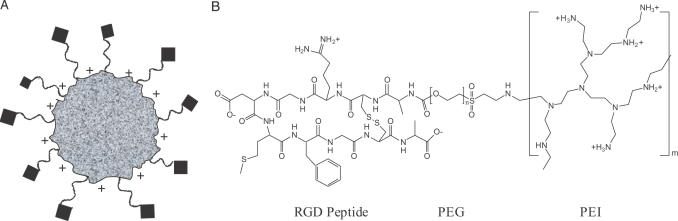
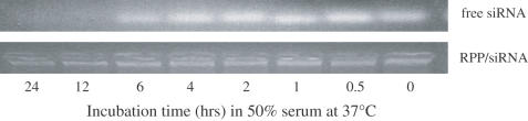
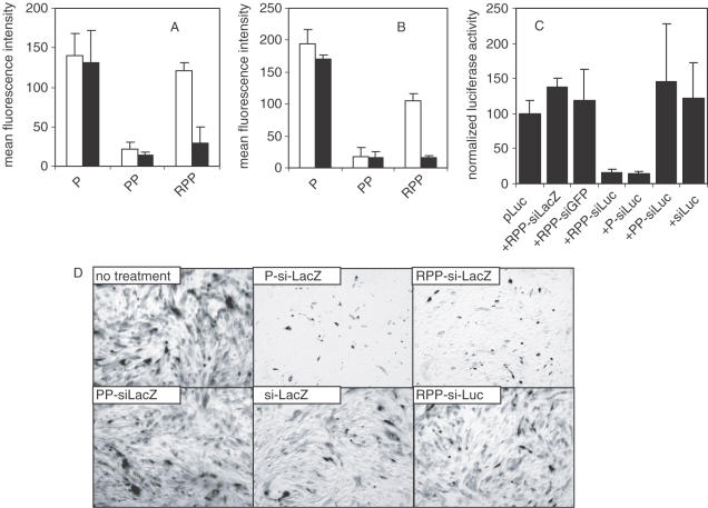
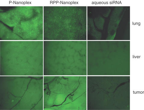
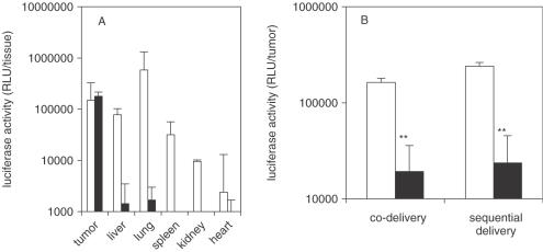
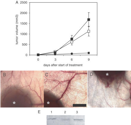
Potent sequence selective gene inhibition by siRNA 'targeted' therapeutics promises the ultimate level of specificity, but siRNA therapeutics is hindered by poor intracellular uptake, limited blood stability and non-specific immune stimulation. To address these problems, ligand-targeted, sterically stabilized nanoparticles have been adapted for siRNA. Self-assembling nanoparticles with siRNA were constructed with polyethyleneimine (PEI) that is PEGylated with an Arg-Gly-Asp (RGD) peptide ligand attached at the distal end of the polyethylene glycol (PEG), as a means to target tumor neovasculature expressing integrins and used to deliver siRNA inhibiting vascular endothelial growth factor receptor-2 (VEGF R2) expression and thereby tumor angiogenesis. Cell delivery and activity of PEGylated PEI was found to be siRNA sequence specific and depend on the presence of peptide ligand and could be competed by free peptide. Intravenous administration into tumor-bearing mice gave selective tumor uptake, siRNA sequence-specific inhibition of protein expression within the tumor and inhibition of both tumor angiogenesis and growth rate. The results suggest achievement of two levels of targeting: tumor tissue selective delivery via the nanoparticle ligand and gene pathway selectivity via the siRNA oligonucleotide. This opens the door for better targeted therapeutics with both tissue and gene selectivity, also to improve targeted therapies with less than ideal therapeutic targets.


Figures

Figure 1
Figure 1
Design of targeted self-assembling siRNA nanoplex and activity in vitro. (A) Schematic structure of the targeted self-assembling nanoplex. Electrostatic interactions between negatively charged siRNA and cationic polymer result in the formation of a complex (speckled particle) that is protected by exposed steric polymer strands (wavy lines), and which gains target selectivity through a targeting ligand moiety coupled to the distal end of the steric polymer (diamonds). (B) Schematic structure of the RPP-polymer. The polymer is designed with the three functional domains needed to obtain the targeted self-assembling nanoplex as described in (A). Branched PEI with a molecular weight of ∼25 kDa (∼581 monomer units) is used as the cationic complexing polymer and contains on average 25% primary, 50% secondary and 25% tertiary amines. Approximately 7% of the amines are modified with PEG with an average molecular weight of 3.4 kDa (80 monomer units). The PEG is conjugated to a folded RGD peptide containing a disulfide bridge between two cysteine residues, a ligand for integrins.
Figure 2
Figure 2
Degradation of siRNA in serum. Degradation of siRNA oligonucleotides when exposed to serum was measured for RPP-nanoplexes and compared with aqueous siRNA. The siRNA nanoplex, or aqueous siRNA, was incubated in 50% serum from 0 to 24 h and then the remaining intact siRNA determined by gel electrophoresis. Staining of the siRNA bound in nanoplexes shows it remains in the loading well without evidence for degradation, while the aqueous siRNA runs as a brightly stained band that diminishes with incubation time.
Figure 3
Figure 3
In vitro activity of siRNA nanoplex. (A and B) siRNA nanoplex binding to cells. 1 × 10⁵ HUVEC (A, open bars) or N2A cells (B, open bars) were incubated with 2 μg fluorescently labeled siRNA formulated in P-, PP- or RPP-nanoplexes for 1 h at 4°C. Both cell types express integrins. After the incubation period, the cells were washed, fixed with 4% buffered formaldehyde and cell-bound fluorescence analyzed by FACS analysis. Unshielded positively charged P-nanoplexes showed relatively high cell binding to both cell types. Shielding of the charged nanoplex with PEG (PP-nanoplexes) reduced cell interaction, which was restored by coupling of the RGD peptide to the distal end of the PEG-shield (RPP-nanoplexes). Pre-incubation of HUVEC (A, closed bars) or N2A (B, closed bars) with a 100-fold molar excess of RGD peptide reduced binding of RPP-nanoplexes while leaving binding of P-or PP-nanoplexes unaffected, indicating that the binding of RPP-nanoplexes to cells is mediated by the RGD peptide targeting ligand. (C) Luciferase silencing in vitro. N2A cells were transfected with 2 μg luciferase plasmid, using cationic lipids without siRNA or cotransfected with 1 μg siRNA formulated as RPP-siLacZ, RPP-siGFP, RPP-siLuc, P-siLuc, PP-siLuc or free siLuc. Sequence-specific silencing of luciferase expression with siLuc is observed for RPP- and P-nanoplexes. Luciferase activity of cells treated with various agents were normalized assuming the activity of cells transfected with luciferase plasmid to be 100%. (D) β-Galactosidase silencing in vitro. SVR-bag 4 endothelial cells, constitutively expressing β-galactosidase were left untreated or incubated with 10 μg of siRNA as follows: P-siLacZ, RPP-siLacZ, PP- siLacZ, RPP-siLuc or free siLacZ. After 3 h incubation, the cells were washed and 48 h after transfection, the cells were stained for β-galactosidase activity. Only panels P-siLacZ and RPP-siLacZ show clear inhibition of β-galactosidase expression.
Figure 4
Figure 4
Tissue distribution of siRNA nanoplexes in N2A tumor-bearing mice. Mice received 40 μg fluorescently labeled siRNA by intravenous injection as P-nanoplexes (left column), or RPP-nanoplexes (middle column) or aqueous solution (right column). One hour after injection, tissues were dissected and examined on a fluorescence microscope. Pictures were taken at equal exposure times for each tissue. P-nanoplexes show punctate fluorescence in all organs, especially high in lung and liver. RPP-nanoplexes show lower fluorescence levels in lung tissue with a punctate distribution and lower non-punctate fluorescence in liver. Higher levels of fluorescence were observed in the tumor as compared with P-nanoplexes. Fluorescence levels after administration of free siRNA were much lower in all organs, as compared with either of the two nanoplex formulations.
Figure 5
Figure 5
Inhibition of plasmid-mediated reporter gene expression in vivo by siRNA-nanoplexes. (A) RPP-plasmid mediated luciferase expression in tumor. N2A-tumor-bearing mice received a single intravenous injection of 40 μg pLuc in either P-nanoplexes (open bars) or RPP-nanoplexes (closed bars). After 24 h following administration, tissues were dissected, homogenized and assayed for luciferase activity. The tissues obtained from mice treated with plasmid in P-nanoplexes show no tumor-specific expression and injection was associated with side effects. Tissues obtained from mice treated with plasmid in RPP-nanoplexes displayed appreciable levels of luciferase expression only in the tumor (n = 5). (B) Plasmid delivery combined with siRNA delivery. N2A-tumor-bearing mice were treated (i.v. injection) with pLuc plasmid as in 5A but received either 13 μg siRNA simultaneously (co-delivery) or 40 μg siRNA 2 h later (sequential delivery) in RPP-nanoplexes. In both the cases, expression of luciferase in the tumor treated with irrelevant siRNA (open bars) was similar to the expression obtained after injection of plasmid alone (A) and treatment with Luc siRNA in RPP-nanoplexes results in ∼90% gene silencing (closed bars) (n = 5).
Figure 6
Figure 6
Tumor growth inhibition by VEGF R2 siRNA nanoplexes. (A) Tumor growth inhibition by siRNA RPP-nanoplexes. Mice were inoculated with N2A tumor cells and left untreated (open squares) or treated every 3 days by tail vein injection with RPP-nanoplexes with siLacZ (filled squares) or siVEGF R2 (filled circles) at a dose of 40 μg per mouse. Treatment was started at the time-point that the tumors became palpable (∼20 mm3). Only VEGF R2-sequence-specific siRNA inhibited tumor growth, whereas treatment with LacZ siRNA did not affect tumor growth rate as compared with untreated controls (n = 5). (B–D) Neovascularization in tumors treated with siRNA RPP-nanoplexes. Representative tumors excised at the end of the tumor growth inhibition experiment (A) were examined using low magnification light microscopy. Trans-illumination of tumor and surrounding skin tissue shows strong neovascularization in mice left untreated (B) and mice treated with RPP-nanoplexes with siRNA-LacZ (C). In contrast, mice treated with RPP-nanoplexes with VEGF R2 siRNA showed low neovascularization and erratic branching of blood vessels (D). Asterisks indicate tumor tissue. Bar = 2 mm. (E) VEGF R2 expression in tumor tissue after treatment with siRNA RPP-nanoplexes. Representative tumors removed at the end of the tumor growth inhibition experiment (A) were homogenized and VEGF R2-expression levels measured by western blotting. The largest tumors in the group treated with mVEGF R2 siRNA in RPP-nanoplexes and the smallest tumors in the other two groups were removed at the end of the tumor growth study 3 days following the final treatment. Lane 1 is untreated tumor, Lane 2 is VEGF R2 siRNA treated tumor and Lane 3 is LacZ siRNA treated tumor.

References

    1. Ferrara N., Hillan,K.J., Gerber,H.-P. and Novotny,W. (2004) Discovery and development of bevacizumab, an anti-VEGF antibody for treating cancer. Nature Rev. Drug Discov., 3, 391–400. - PubMed
    1. Elbashir S.M., Harborth,J., Weber,K. and Tuschl,T. (2002) Analysis of gene function in somatic mammalian cells using small interfering RNAs. Methods, 26, 199–213. - PubMed
    1. Paddison P.J. and Hannon,G.J. (2003) siRNAs and shRNAs: skeleton keys to the human genome. Curr. Opin. Mol. Ther., 5, 217–224. - PubMed
    1. Carpenter A.E. and Sabatini,D.M. (2004) Systematic genome-wide screens of gene function. Nature Rev. Genet., 5, 11–22. - PubMed
    1. Ganju P. and Hall,J. (2004) Potential applications of siRNA for pain therapy. Expert Opin. Biol. Ther., 4, 531–542. - PubMed

Publication types

MeSH terms