

Evaluation of hypoxia in an experimental rat tumour model by [(18)F]fluoromisonidazole PET and immunohistochemistry
- PMID: 15520822
- PMCID: PMC2409764
- DOI: 10.1038/sj.bjc.6602219
Evaluation of hypoxia in an experimental rat tumour model by [(18)F]fluoromisonidazole PET and immunohistochemistry
Abstract
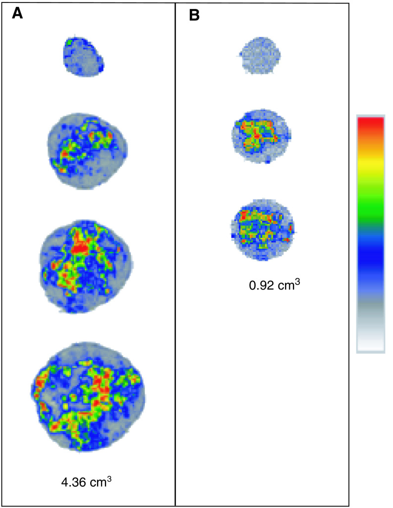
This study aimed to evaluate tumour hypoxia by comparing [(18)F]Fluoromisonidazole uptake measured using positron emission tomography ([(18)F]FMISO-PET) with immunohistochemical (IHC) staining techniques. Syngeneic rhabdomyosarcoma (R1) tumour pieces were transplanted subcutaneously in the flanks of WAG/Rij rats. Tumours were analysed at volumes between 0.9 and 7.3 cm(3). Hypoxic volumes were defined using a 3D region of interest on 2 h postinjection [(18)F]FMISO-PET images, applying different thresholds (1.2-3.0). Monoclonal antibodies to pimonidazole (PIMO) and carbonic anhydrase IX (CA IX), exogenous and endogenous markers of hypoxia, respectively, were used for IHC staining. Marker-positive fractions were microscopically measured for each tumour, and hypoxic volumes were calculated. A heterogeneous distribution of hypoxia was observed both with histology and [(18)F]FMISO autoradiography. A statistically significant correlation (P<0.05) was obtained between the hypoxic volumes defined with [(18)F]FMISO-PET and the volumes derived from the PIMO-stained tumour sections (r=0.9066; P=0.0001), regardless of the selected threshold between 1.4 and 2.2. A similar observation was made with the CA IX staining (r=0.8636; P=0.0006). The relationship found between [(18)F]FMISO-PET and PIMO- and additionally CA IX-derived hypoxic volumes in rat rhabdomyosarcomas indicates the value of the noninvasive imaging method to measure hypoxia in whole tumours.
Figures
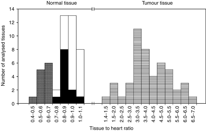
 , muscle (n=24; that is, front leg muscle n=12 and hind leg muscle n=12) □ and a body area 15 mm above the heart (n=12) ▪ on 12 randomly chosen 2 h p.i. images. Similarly, tumours (n=48)
  were analysed on 2 h p.i. images. For all the tissues, cumulative histogram analysis was carried out.
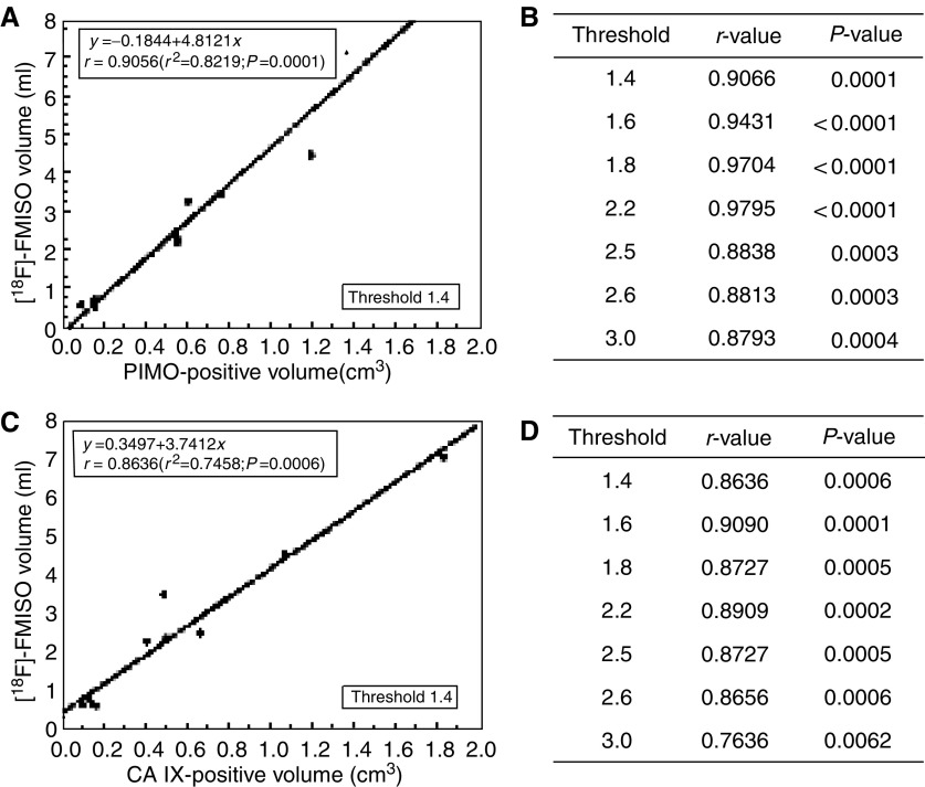
References
-
- Airley RE, Loncaster J, Raleigh JA, Harris AL, Davidson SE, Hunter RD, West CML, Stratford IJ (2003) Glut-1 and CA IX as intrinsic markers of hypoxia in carcinoma of the cervix: relationship to pimonidazole binding. Int J Cancer 104: 85–91, doi: 10.1002/ijc.10904 - PubMed
-
- Ballinger JR (2001) Imaging hypoxia in tumors. Semin Nucl Med 4: 321–329, doi: 10.1053/snuc.2001.26191 - PubMed
-
- Bentzen L, Keiding S, Horsman M, Falborg L, Hansen SB, Overgaard J (2000) Feasibility of detecting hypoxia in experimental mouse tumours with 18F-fluorinated tracers and Positron Emission Tomography – a study evaluating [18F]Fluoromisonidazole and [18F]Fluoro-2-deoxy-D-glucose. Acta Oncol 39: 629–637 - PubMed
-
- Bentzen L, Keiding S, Horsman MR, Grönroos T, Hansen SB, Overgaard J (2002) Assessment of hypoxia in experimental mice tumours by [18F]Fluoromisonidazole PET and pO2 electrode measurements – Influence of tumour volume and carbogen breathing. Acta Oncol 41: 304–312 - PubMed
MeSH terms
Substances
LinkOut - more resources
Full Text Sources
Other Literature Sources
