. 2004 Nov;114(9):1260-71.
doi: 10.1172/JCI21378.

Semaphorin 3F, a chemorepulsant for endothelial cells, induces a poorly vascularized, encapsulated, nonmetastatic tumor phenotype

Affiliations

Semaphorin 3F, a chemorepulsant for endothelial cells, induces a poorly vascularized, encapsulated, nonmetastatic tumor phenotype

Diane R Bielenberg et al. J Clin Invest. 2004 Nov.

Abstract

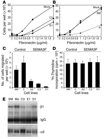
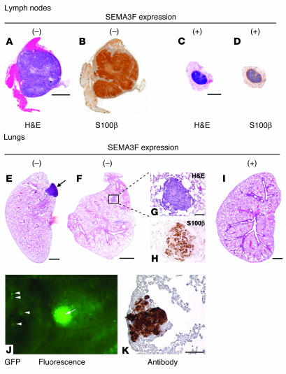
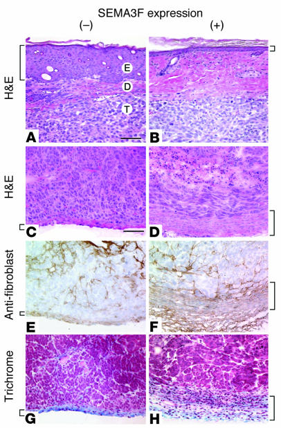
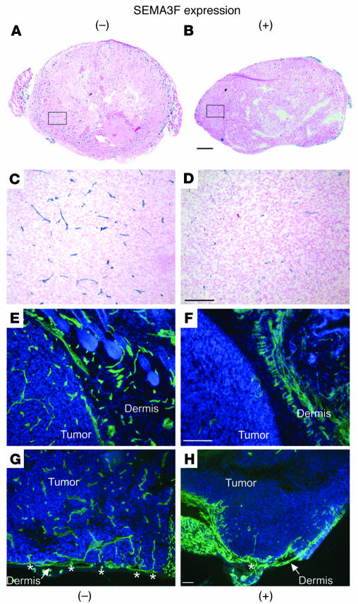
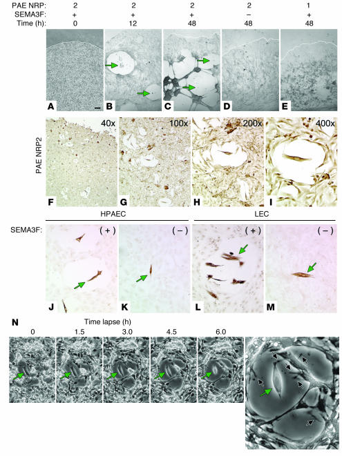
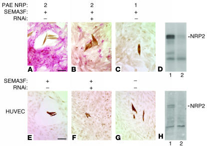
Melanoma is the most lethal skin cancer. Most deaths from melanoma result from metastases. Semaphorins have been shown to inhibit neuronal and endothelial cell migration, but the effects of semaphorins on tumor metastasis have not been documented. We found that semaphorin 3F (SEMA3F) was markedly downregulated in highly metastatic human cell lines in vitro and in vivo, which suggested that it may be a metastasis inhibitor. Metastatic human melanoma cells were transfected with SEMA3F and implanted into mice; the resultant tumors did not metastasize. Rather, the primary tumors resembled benign nevi characterized by large areas of apoptosis, diminished vascularity, inhibition of hyperplasia in overlying epidermal cells, and encapsulated tumor borders delineated by thick layers of fibroblasts and collagen matrix. This phenotype is in stark contrast to highly invasive, vascular mock-transfected tumors. In vitro, tumor cells expressing SEMA3F had a diminished capacity to adhere and migrate on fibronectin. Consistent with semaphorin-mediated chemorepulsion of neurons, tumor cells expressing SEMA3F were chemorepulsive for vascular and lymphatic endothelial cells expressing neuropilin-2 (NRP2), a novel mechanism for a tumor angiogenesis inhibitor. The repulsive activity was abrogated by NRP2 RNA interference. Together these results indicate that SEMA3F is a potent metastasis inhibitor that targets both tumor and stromal cells and raise the possibility of SEMA3F having therapeutic potential.


Figures

Figure 1
Figure 1
Metastatic human cell lines downregulate SEMA3F. (A) Northern blot analysis of paired human cancer cell lines with low (–) versus high (+) metastatic potential. The paired cell lines include prostate carcinoma cells PC3M (–) versus PC3MLN4 (+), transitional cell bladder carcinoma cells 253J (–) versus 253JBV (+), and melanoma cells A375P (–) versus SM (+). mRNA expression analysis of SEMA3F, NRP1, and NRP2 is demonstrated. (B) RT-PCR analysis for VEGFRs in SM cells and HUVECs. GAPDH was used as a control. (C) Western blot analysis of NRP1 and NRP2 in SM cells and various ECs including LECs, PAE NRP2 cells, PAE NRP1 cells, HUVECs, and HPAECs. (D) Northern blot analysis (top panel) and Western blot analysis (bottom panel) of myc-tagged SEMA3F expression in various transfected clones in vitro. (E) Northern blot analysis of representative tumor clones in vivo. mRNA was isolated directly from tumors grown in mice for 4–5 weeks and blotted for expression of SEMA3F and NRP2. Actin is used as a loading control in A, D (top), and E. SM, (untransfected) A375SM cells; Mo, mock transfected A375SM cells containing empty vector only; H10, clone transfected with the SEMA3F plasmid that did not express detectable SEMA3F.
Figure 2
Figure 2
SEMA3F inhibits adhesion and migration, but not proliferation, of tumor cells. (A) Adhesion of mock-transfected SM cells (squares) and 2 SM/SEMA3F clones, C3 (circles) and E1 (triangles), to FN. (B) Adhesion of parental (A375P) nonmetastatic melanoma cells (triangles), SM metastatic cells (circles), and mock-transfected SM cells (squares) to FN. (C) Boyden chamber motility assay of representative transfected clones. (D) 3H-thymidine incorporation into DNA to measure proliferation in representative transfected clones. (E) Western blot analysis of integrins β1 and α5 in 3 representative SEMA3F-transfected clones (C3, E1, and D1) following immunoprecipitation with α5β1 antibody.
Figure 3
Figure 3
SEMA3F overexpression results in tumor apoptosis. Scans of whole-tumor cross sections. (AC) ISH for SEMA3F expression (dark purple). (DF) H&E staining. (GI) TUNEL staining to detect apoptotic cells (brown). (JL) PCNA staining to detect dividing cells (brown). Arrows denote areas of apoptosis that lack SEMA3F or PCNA staining. Scale bars: 2 mm.
Figure 4
Figure 4
SEMA3F inhibits lymph node and lung metastasis. (AD) Scans of lymph nodes stained with H&E (A and C) or the melanoma marker S100β (B and D, brown). Scale bars: 1 mm. (E, F, and I) Scans of lung sections stained by H&E. Scale bars: 1 mm. The arrow points to a macrometastasis (E), and the box surrounds a micrometastasis (F). (G and H) The micrometastasis in F is shown magnified and stained with H&E (G) and S100β (H, brown). Scale bar: 100 μm. No metastases were detected in lungs of mice bearing SM/SEMA3F tumors (I). (J and K) Use of GFP-infected cells to detect lung metastases. Large metastasis (J, arrow) as well as small metastases (J, arrowheads) fluoresced green in lungs from mice with control tumors. IHC with antibodies to GFP also detected micrometastases in lungs from mice with control tumors (K, brown). Scale bar: 100 μm.
Figure 5
Figure 5
SEMA3F expression alters the tumor microenvironment. (A and B) H&E staining of tumors with and without SEMA3F expression and overlying skin. E, epidermis; D, dermis; T, tumor. Scale bar: 100 μm. Mouse epidermal cells overlying control tumors were hyperplastic (A, bracket), while the epidermis overlying tumors expressing SEMA3F remained at nearly normal thickness (B, bracket). (CH) Tumor/stromal borders, with and without SEMA3F expression. (C and D) H&E. (E and F) IHC using antibodies to fibroblasts (brown). (G and H) Trichrome staining; collagen is stained in blue. (C, E, and G) Tumors without SEMA3F had nondelineated borders and invasive tumor leading edges. (D, F, and H) Tumors expressing SEMA3F were surrounded by thick layers of stromal cells (brackets) forming a capsule around the tumor. The capsule was composed largely of fibroblasts (F) that deposited collagen matrix (H). Scale bar: 100 μm.
Figure 6
Figure 6
SEMA3F inhibits tumor angiogenesis. CD31 staining in tumors lacking (A, C, E, and G) and expressing SEMA3F (B, D, F, and H). Vessels appear blue in thin (8 μm) cryosections (AD) or fluorescent green in thick (30 μm) cryosections (EH). (A and B) Scans of whole-primary-tumor cross sections stained with CD31. Scale bar: 1 mm. (C and D) A representative field from each tumor (in a non-necrotic area) is shown at higher magnification (C from box in A; D from box in B). (EH) Thick cryosections were used to detect neovascularization and sprouting (asterisks) from surrounding skin vessels into the tumors. Scale bars in CH: 200 μm.
Figure 7
Figure 7
SEMA3F-induced EC chemorepulsion. (AE) PAE NRP2 (AD) or PAE NRP1 cells (E) were cultured to confluence. At time 0, SM/SEMA3F cells were added into the culture (A). EC retraction was apparent at 12 hours (B) and was even more dramatic at 48 hours (C). Green arrows point to the SM/SEMA3F cells at the center of the clearance zones. No clearance zone was observed when SM cells lacking SEMA3F were added (D) or when PAE NRP1 cells were used (E). Scale bar: 200 μm. (FI) The repulsion of PAE NRP2 cells by SM/SEMA3F cells (brown color due to melanoma marker S100β staining) at 9 hours of coculture is shown at various magnifications – 40× (F), 100× (G), 200× (H), and 400× (I) – to demonstrate the large number of clearance zones observable at low magnification and the clearance zone induced by 1 individual SM/SEMA3F cell at high magnification. (JM) The repulsion of primary ECs. HPAECs (J) and LECs (L) are repelled from SM/SEMA3F cells, but HPAECs (K) and LECs (M) are not repelled from control SM/Mock cells. Representative tumor cells are denoted by green arrows. Scale bars: 100 μm. (N) Time-lapse photography and video (see supplemental data) of the interaction of SM/SEMA3F and PAE NRP2 cells. The central tumor cell (green arrow) is expressing SEMA3F, and the surrounding cells are PAE cells expressing NRP2. Notice the ECs moving away from the source of SEMA3F over time, creating a clearance zone. In the video and in a higher-magnification view (right panel), the ECs can be seen to extend and retract numerous filopodia (black arrows) as they sense their environment.
Figure 8
Figure 8
EC repulsion by SEMA3F is NRP2 dependent. (A and B) PAE NRP2 cells are stained pink for NRP2 protein. SM/SEMA3F cells (brown) induce clearance zones (A), but not in cells treated with RNAi (B). (C) No clearance zones are found in PAE NRP1 cells. (D) Western blot analysis of PAE NRP2 cells with and without RNAi. PAE NRP2/RNAi cells express dramatically reduced NRP2 protein (B; and D, lane 2). (EH) HUVECs. SM/SEMA3F cells induce a zone of clearance (E), but not in the presence of RNAi (F) or the absence of SEMA3F (G). (H) Western blot analysis of HUVECs with and without RNAi. HUVECs/RNAi cells express dramatically reduced NRP2 protein (H, lane 2). Scale bars: 100 μm.

References

    1. Kolodkin AL, et al. Neuropilin is a semaphorin III receptor. Cell. 1997;90:753–762. - PubMed
    1. Soker S, Takashima S, Miao HQ, Neufeld G, Klagsbrun M. Neuropilin-1 is expressed by endothelial and tumor cells as an isoform-specific receptor for vascular endothelial growth factor. Cell. 1998;92:735–745. - PubMed
    1. Gagnon ML, et al. Identification of a natural soluble neuropilin-1 that binds vascular endothelial growth factor: in vivo expression and antitumor activity. Proc. Natl. Acad. Sci. U. S. A. 2000;97:2573–2578. - PMC - PubMed
    1. Harper J, Gerstenfeld LC, Klagsbrun M. Neuropilin-1 expression in osteogenic cells: down-regulation during differentiation of osteoblasts into osteocytes. J. Cell. Biochem. 2001;81:82–92. - PubMed
    1. Chen H, Chedotal A, He Z, Goodman CS, Tessier-Lavigne M. Neuropilin-2, a novel member of the neuropilin family, is a high affinity receptor for the semaphorins Sema E and Sema IV but not Sema III. Neuron. 1997;19:547–559. - PubMed

Publication types

MeSH terms