. 2004 Nov;114(9):1308-16.
doi: 10.1172/JCI19603.

Imatinib mesylate inhibits the profibrogenic activity of TGF-beta and prevents bleomycin-mediated lung fibrosis

Affiliations

Imatinib mesylate inhibits the profibrogenic activity of TGF-beta and prevents bleomycin-mediated lung fibrosis

Craig E Daniels et al. J Clin Invest. 2004 Nov.

Abstract

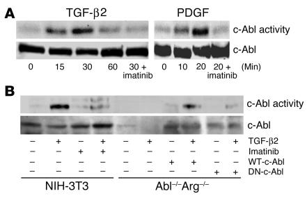
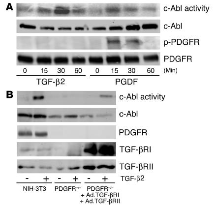
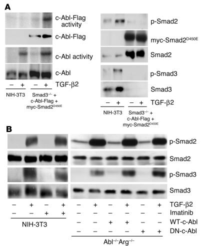
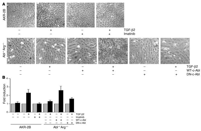
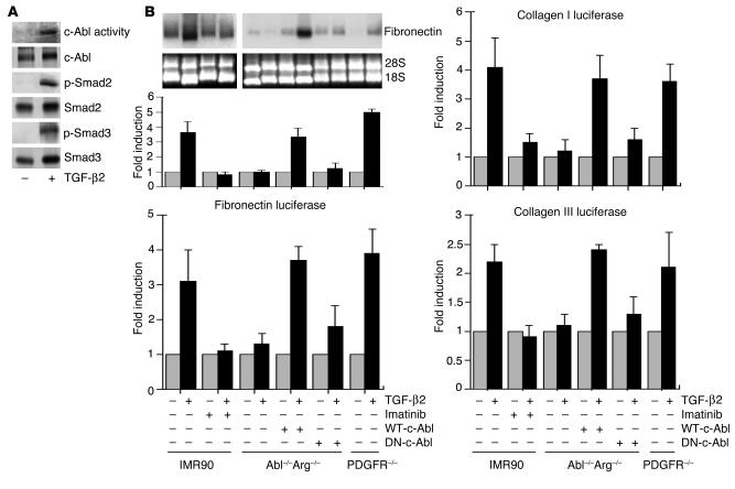
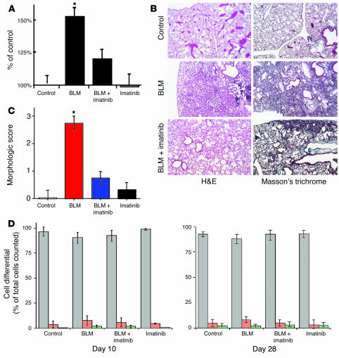
Idiopathic pulmonary fibrosis is a progressive and fatal fibrotic disease of the lungs with unclear etiology. Prior efforts to treat idiopathic pulmonary fibrosis that focused on anti-inflammatory therapy have not proven to be effective. Recent insight suggests that the pathogenesis is mediated through foci of dysregulated fibroblasts driven by profibrotic cytokine signaling. TGF-beta and PDGF are 2 of the most potent of these cytokines. In the current study, we investigated the role of TGF-beta-induced fibrosis mediated by activation of the Abelson (Abl) tyrosine kinase. Our data indicate that fibroblasts respond to TGF-beta by stimulating c-Abl kinase activity independently of Smad2/3 phosphorylation or PDGFR activation. Moreover, inhibition of c-Abl by imatinib prevented TGF-beta-induced ECM gene expression, morphologic transformation, and cell proliferation independently of any effect on Smad signaling. Further, using a mouse model of bleomycin-induced pulmonary fibrosis, we found a significant inhibition of lung fibrosis by imatinib. Thus, Abl family members represent common targets for the modulation of profibrotic cytokine signaling.


Figures

Figure 1
Figure 1
TGF-β2 stimulates c-Abl kinase activity. (A) NIH-3T3 fibroblasts were grown to confluence, placed in 0.1% FBS DMEM overnight, and stimulated with 10 ng/ml TGF-β2 or 25 ng/ml PDGF-AB for the indicated times. Parallel plates were pretreated for 20 minutes with 10 μg/ml imatinib prior to growth factor addition. Following c-Abl immunoprecipitation, in vitro kinase activity was determined as described in Methods. Similar results were obtained with AKR-2B fibroblasts and TGF-β1 (data not shown). Lower half: Prior to immunoprecipitation and kinase assay, 100 μg of protein was used for c-Abl Western analysis. (B) NIH-3T3 cells, Abl–/–Arg–/– MEFs, or Abl–/–Arg–/– fibroblasts stably expressing (+) WT-c-Abl or dominant negative c-Abl (DN-c-Abl) were plated at 2.0 106 cells per 100-mm plate and grown overnight in 20% FBS DMEM. The medium was changed to 0.5% FBS DMEM for 24 hours, and the cultures were left untreated (–) or stimulated (+) with 10 ng/ml TGF-β2 for 30 minutes. Parallel plates were pretreated for 20 minutes with 10 μg/ml imatinib prior to growth factor addition. c-Abl kinase activity (top) and Western analysis (bottom) were determined as in A.
Figure 2
Figure 2
TGF-β2 activation of c-Abl is independent of PDGFR signaling. (A) NIH-3T3 cells were treated for the indicated times with TGF-β2 (10 ng/ml) or PDGF-AB (25 ng/ml). Cell lysates were prepared and processed for c-Abl kinase activity (first panel), total c-Abl protein (second panel), tyrosine-phosphorylated PDGF-β receptor (p-PDGFR; third panel), or total PDGF-β receptor (fourth panel) as described in Methods. (B) NIH-3T3 cells were treated as described in Figure 1. PDGF-α and -β receptor–null F cells were seeded at 1.5 × 10⁶ cells per 100-mm dish. Following overnight growth, the medium was replaced with serum-free DMEM alone (PDGFR–/–) or adenovirus (MOI 100) expressing the type I and type II TGF-β receptors (Ad.TGF-βRI and Ad.TGF-βRII, respectively) for 24 hours. Cultures were left untreated (–) or stimulated (+) with 10 ng/ml TGF-β2 for 30 minutes and processed for c-Abl kinase activity (first panel), or total c-Abl (second panel), PDGF-β receptor (third panel), type I TGF-β receptor (TGF-βRI; fourth panel), or type II TGF-β receptor (TGF-βRII; fifth panel) protein.
Figure 3
Figure 3
TGF-β2 activation of c-Abl signaling and TGF-β2 activation of Smad signaling are independent. (A) Left: Smad3 KO MEFs (Smad3–/–) were transfected with Flag-tagged c-Abl (c-Abl–Flag) and dominant negative Smad2 (myc-Smad2D450E) as described in Methods. NIH-3T3 cells were treated as described in Figure 1. Following 30-minute stimulation in the absence (–) or presence (+) of 10 ng/ml TGF-β2, kinase activity of the transfected (c-Abl–Flag activity) or endogenous (c-Abl activity) c-Abl protein was determined using anti-Flag or K12 serum respectively. The corresponding total protein is indicated in the second and fourth panels. Right: NIH-3T3 or Smad2/3 KO cells were treated with (+) or without (–) TGF-β as described for the left panels. Following 30-minute stimulation, lysates containing equivalent protein were Western-blotted for the indicated proteins. (B) Left: NIH-3T3 cells were treated with (+) or without (–) TGF-β2 and/or imatinib as described in Figure 1. Following 30-minute stimulation, lysates containing equivalent protein were Western-blotted for phospho-Smad2 (p-Smad2), phospho-Smad3 (p-Smad3), or the corresponding total protein. Right: Abl–/–Arg–/– MEFs or Abl–/–Arg–/– fibroblasts stably expressing (+) WT-c-Abl or dominant negative c-Abl were treated with (+) or without (–) TGF-β2 for 30 minutes and processed for the indicated Smad protein as described for the left panels.
Figure 4
Figure 4
c-Abl is required for TGF-β–induced morphologic alteration and cell proliferation. (A) AKR-2B cells, Abl–/–Arg–/– MEFs, or Abl–/–Arg–/– fibroblasts stably expressing (+) WT-c-Abl or dominant negative c-Abl (DN-c-Abl) were grown to confluence as described in Methods. Cultures were stimulated with DMEM alone or supplemented (+) with 5 μg/ml imatinib and/or 10 ng/ml TGF-β2. Following 48-hour incubation, representative areas were photographed at 20 phase. (B) Triplicate plates from A were trypsinized, and cells were counted with a hemocytometer.
Figure 5
Figure 5
TGF-β stimulates c-Abl–dependent signaling. (A) IMR90 cells were treated with DMEM alone (–) or containing 10 ng/ml TGF-β2 (+). Following 30-minute stimulation, c-Abl kinase activity or Smad2 (p-Smad2) and Smad3 (p-Smad3) phosphorylation was determined. Total c-Abl, Smad2, and Smad3 protein is shown below the corresponding activity. (B) Top blots: IMR90 cells, Abl–/–Arg–/– MEFs, Abl–/–Arg–/– fibroblasts stably expressing (+) WT-c-Abl or dominant negative c-Abl, or PDGF-α and -β receptor–null F cells (PDGFR–/–) were left untreated (–) or treated (+) for 24 hours with TGF-β2 (10 ng/ml) and/or imatinib (10 μg/ml). Imatinib was added 20 minutes before TGF-β2, and fibronectin mRNA accumulation was determined. Bottom blots: Ethidium bromide staining for 28S and 18S ribosomal RNA subunits prior to Northern analysis. Top left graph: The fold fibronectin mRNA stimulation by TGF-β2 is indicated for the various cell types. Results represent the mean ± SE of 2 separate experiments. Bottom left graph: Cell lines as described above were transfected with a fibronectin luciferase construct. The fold induction of 10 ng/ml TGF-β2 with or without 5 μg/ml imatinib was determined as described in Methods and by Penheiter et al. (51). Right graphs: Collagen I (top) and collagen III (bottom) luciferase activity was determined as described for the bottom left graph. Results for all luciferase assays represent the mean ± SE of 2 separate experiments, each done in triplicate.
Figure 6
Figure 6
Imatinib reduces hydroxyproline content and prevents histopathologic changes in BLM-treated lungs. (A) Twenty-eight days after intratracheal instillation of BLM or saline, mice were sacrificed (control, n = 12; BLM, n = 15; BLM plus imatinib, n = 14; imatinib, n = 8). The left lung of each animal was used for hydroxyproline assay as described in Methods and previously (52). *P < 0.05 vs. BLM mice receiving imatinib. There was no significant difference (P > 0.05) among control, BLM plus imatinib, and imatinib groups. (B) Mice were treated as in A. The right lung was sectioned and stained with H&E or Masson’s trichrome to visualize collagen. Representative images are shown at ×10 phase. (C) A morphologic score (44) was assigned in a blinded fashion to H&E-stained samples. Zero indicates no fibrosis, 1 indicates occasional small subpleural foci, 2 indicates interalveolar septal thickening and subpleural foci, and 3 indicates continuous interalveolar and subpleural fibrosis. There was no significant difference among the control, BLM plus imatinib, and imatinib groups. (D) Mice were treated as described for A. Immediately prior to explantation trachea were cannulated and lavaged with a total of 10 ml 0.9 normal saline. The collected fluid was centrifuged, and the cell differential was determined. Gray bars indicate percent macrophages, while red and green reflect polymorphonuclear cells and lymphocytes, respectively. Error bars indicate the SE of measurement between samples within the same group.

References

    1. Idiopathic pulmonary fibrosis: diagnosis and treatment. International consensus statement. American Thoracic Society (ATS), and the European Respiratory Society (ERS) Am. J. Respir. Crit. Care Med. 2000;161:646–664. - PubMed
    1. Coultas DB, Sumwalt RE, Black WC, Sobonya RE. The epidemiology of interstitial lung diseases. Am. J. Respir. Crit. Care Med. 1994;150:967–972. - PubMed
    1. Bjoraker JA, et al. Prognostic significance of histopathologic subsets in idiopathic pulmonary fibrosis. Am. J. Respir. Crit. Care Med. 1998;157:199–203. - PubMed
    1. Strieter RM. Mechanisms of pulmonary fibrosis: conference summary. Chest. 2001;120(1 Suppl.):77S–85S. - PubMed
    1. Selman M, King TE, Pardo A. Idiopathic pulmonary fibrosis: prevailing and evolving hypotheses about its pathogenesis and implications for therapy. Ann. Intern. Med. 2001;134:136–151. - PubMed

Publication types

MeSH terms