. 2004 Nov;114(9):1334-42.
doi: 10.1172/JCI22608.

Recruitment and expansion of dendritic cells in vivo potentiate the immunogenicity of plasmid DNA vaccines

Affiliations

Recruitment and expansion of dendritic cells in vivo potentiate the immunogenicity of plasmid DNA vaccines

Shawn M Sumida et al. J Clin Invest. 2004 Nov.

Abstract

DCs are critical for priming adaptive immune responses to foreign antigens. However, the utility of harnessing these cells in vivo to optimize the immunogenicity of vaccines has not been fully explored. Here we investigate a novel vaccine approach that involves delivering synergistic signals that both recruit and expand DC populations at the site of antigen production. Intramuscular injection of an unadjuvanted HIV-1 envelope (env) DNA vaccine recruited few DCs to the injection site and elicited low-frequency, env-specific immune responses in mice. Coadministration of plasmids encoding the chemokine macrophage inflammatory protein-1alpha (MIP-1alpha) and the DC-specific growth factor fms-like tyrosine kinase 3 ligand with the DNA vaccine resulted in the recruitment, expansion, and activation of large numbers of DCs at the site of inoculation. Consistent with these findings, coadministration of these plasmid cytokines also markedly augmented DNA vaccine---elicited cellular and humoral immune responses and increased protective efficacy against challenge with recombinant vaccinia virus. These data suggest that the availability of mature DCs at the site of inoculation is a critical rate-limiting factor for DNA vaccine immunogenicity. Synergistic recruitment and expansion of DCs in vivo may prove a practical strategy for overcoming this limitation and potentiating immune responses to vaccines as well as other immunotherapeutic strategies.


Figures

Figure 1
Figure 1
Histopathology of injection sites. BALB/c mice (n = 4 per group) were immunized intramuscularly with (A) saline; (B) gp120 DNA vaccine alone; or gp120 DNA vaccine with (C) plasmid Flt3L, (D) plasmid MIP-1α, or (E) both plasmid Flt3L and plasmid MIP-1α. We injected 50 μg of each plasmid with sufficient sham plasmid to keep the total DNA dose per mouse constant. Muscle sections (5 μm thick) were stained with H&E on day 7 following immunization. Magnification, ×20.
Figure 2
Figure 2
Immunohistochemistry of injection sites. The muscle sections (5 μm thick) from the vaccinated mice described in Figure 1 were stained with mAbs specific for murine (A) CD3, (B) CD11b, (C) S100, (D) CD83, (E) MHC class II, (F) CD80, or (G) and isotype control. Magnification, ×20.
Figure 3
Figure 3
Analysis of DCs extracted from injected muscles. BALB/c mice were immunized as described in Figure 1. On day 7 following immunization, muscles were excised, homogenized, and digested with collagenase and trypsin (n = 8 per group). Cell suspensions were analyzed by 4-color flow cytometry, and DCs were defined as gated CD3CD19 class II+CD11c+ cells. (A) Mean total number of extracted cells and (B) mean total number of extracted DCs and CD80hi DCs per muscle are shown. (C) Percentage of total extracted cells that were DCs and (D) percentage of DCs that were CD80hi are also shown. In all samples, less than 5% of the cells were CD3+ or CD19+ lymphocytes.
Figure 4
Figure 4
Immunogenicity of MIP-1α/Flt3L–augmented DNA vaccines. BALB/c mice (n = 8 per group) were immunized with sham plasmid; the gp120 DNA vaccine alone; or the gp120 DNA vaccine with plasmid Flt3L, plasmid MIP-1α, or the combination of both plasmid cytokines. We injected 50 μg of each plasmid with sufficient sham plasmid to keep the total DNA dose per mouse constant. Vaccine-elicited immune responses were assessed by (A) Dd/P18 tetramer binding to CD8+ T lymphocytes, (B) env-pooled peptide and P18 epitope peptide-specific ELISPOT assays, and (C) gp120-specific ELISAs. The ELISPOT and ELISA assays were performed on day 28 following immunization. SFCs, spot forming cells.
Figure 5
Figure 5
Generalizability of MIP-1α/Flt3L–augmented DNA vaccines. (A) BALB/c mice or C57BL/6 mice were immunized, respectively, with 50 μg HIV env gp120 DNA vaccine or 50 μg SIV gag DNA vaccine, each with or without plasmid MIP-1α and plasmid Flt3L. ELISPOT assays were performed using pooled env peptides and the P18 epitope peptide for the env-vaccinated mice, or pooled gag peptides and the AL11 epitope peptide for the gag-vaccinated mice. (B) ELISPOT assays were performed using splenocytes from env-vaccinated BALB/c mice depleted of CD4+ or CD8+ T lymphocytes.
Figure 6
Figure 6
Secondary responses following cytokine-augmented DNA vaccine priming and DNA vaccine booster immunization. BALB/c mice (n = 4 per group) were primed with 50 μg gp120 DNA vaccine with or without (A) plasmid MIP-1α and plasmid Flt3L or (B) plasmid MIP-1α and plasmid CD40L. At week 6 following immunization, all mice were given booster immunizations of 50 μg gp120 DNA vaccine. Vaccine-elicited cellular immune responses were assessed by Dd/P18 tetramer binding to CD8+ T lymphocytes following the booster immunization.
Figure 7
Figure 7
Mechanistic studies for plasmid MIP-1α and plasmid Flt3L. (A) BALB/c mice were immunized intramuscularly with sham plasmid, gp120 DNA vaccine alone, gp120 DNA vaccine mixed with plasmid MIP-1α and plasmid Flt3L (delivered equally in both legs), or gp120 DNA vaccine in the left leg (L) and plasmid MIP-1α and plasmid Flt3L in the right leg (R). (B) Mice were immunized with the gp120 DNA vaccine, with or without plasmid MIP-1α and plasmid Flt3L, and received daily i.v. and i.p. injections of saline, 1 μg human MIP-1α protein (hu MIP-1α), or 1 μg murine MIP-1α protein (mu MIP-1α) for 3 days. ND, not done. (C) Mice were immunized with the gp120 DNA vaccine with or without plasmid MIP-1α and plasmid Flt3L at doses of 50 μg, 5 μg, or 0.5 μg of each plasmid in 50-μl injection volumes. (D) Mice were immunized with the gp120 DNA vaccine with or without plasmid MIP-1α and plasmid Flt3L at doses of 50 μg of each plasmid in 50-μl or 15-μl injection volumes. Vaccine-elicited cellular immune responses were assessed by Dd/P18 tetramer binding to CD8+ T lymphocytes on day 10 following immunization.
Figure 8
Figure 8
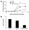
Recombinant vaccinia virus challenge. BALB/c mice (n = 4 per group) were immunized intramuscularly with sham plasmid, gp120 DNA vaccine, or gp120 DNA vaccine with plasmid Flt3L and plasmid MIP-1α. At week 12 following immunization, mice were challenged i.p. with 107 PFU recombinant vaccinia virus expressing HIV-1 env IIIB. (A) Anamnestic immune responses were assessed by Dd/P18 tetramer binding to CD8+ T lymphocytes following challenge. (B) Vaccinia virus titers (PFU) were assessed in ovaries harvested on day 7 following challenge.

Comment in

References

    1. Banchereau J, Steinman RM. Dendritic cells and the control of immunity. Nature. 1998;392:245–252. - PubMed
    1. Mellman I, Steinman RM. Dendritic cells: specialized and regulated antigen processing machines. Cell. 2001;106:255–258. - PubMed
    1. Wolff JA, et al. Direct gene transfer into mouse muscle in vivo. Science. 1990;247:1465–1468. - PubMed
    1. Donnelly JJ, Ulmer JB, Shiver JW, Liu MA. DNA vaccines. Annu. Rev. Immunol. 1997;15:617–648. - PubMed
    1. Calarota S, et al. Cellular cytotoxic response induced by DNA vaccination in HIV-1–infected patients. Lancet. 1998;351:1320–1325. - PubMed

Publication types

MeSH terms