

Testosterone increases bone mineral density in female-to-male transsexuals: a case series of 15 subjects
- PMID: 15521957
- PMCID: PMC3098904
- DOI: 10.1111/j.1365-2265.2004.02125.x
Testosterone increases bone mineral density in female-to-male transsexuals: a case series of 15 subjects
Abstract
Objective: Testosterone therapy for osteoporosis has not been studied extensively in women because of its potential to cause virilization. Female-to-male transsexuals are genetic females who suffer from gender dysphoria and thus take supra-physiologic doses of testosterone to change from the female to male phenotype. The aim of this study is to examine the effects of testosterone treatment on the genetic female skeleton.
Patients and design: A group of 15 female-to-male transsexuals was prospectively enrolled for observation over a 2-year period. The subjects had a mean age of 37.0 +/- 3.0 years. All of the subjects self-administered testosterone esters intramuscularly at a mean dose of 70.7 +/- 4.5 mg weekly.
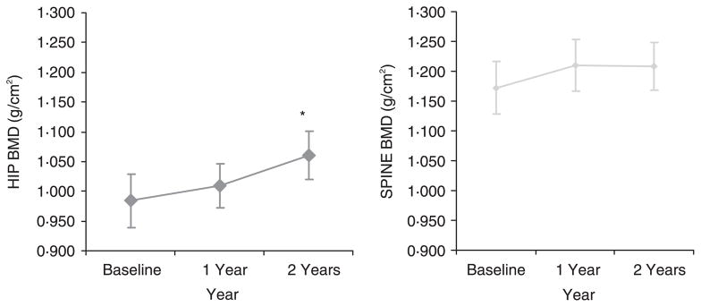
Measurements: The subjects had measurements of bone mineral density (BMD) by dual X-ray absorptiometry (DXA) of the femoral neck and spine (L2-L4) at 12-month intervals. They had determinations of serum oestradiol, testosterone, soluble RANKL (sRANKL), osteoprotegerin (OPG) and urine N-telopeptide (NTX) at the date of enrolment and at the end of 2 years. results There was a significant positive increase in mean BMD of 7.8% at the femoral neck and a nonsignificant increase in mean BMD of 3.1% at the spine over 2 years. The levels of testosterone reached the upper normal range for males and the levels of oestradiol declined to near the postmenopausal range. sRANKL levels decreased significantly in female-to-male transsexuals who newly initiated testosterone therapy. There was no significant change in urine NTX or serum OPG during the study.
Conclusions: We conclude that supra-physiologic testosterone therapy increases BMD at the hip while maintaining BMD at the spine in female-to-male transsexuals. The effects of testosterone may be the result of testosterone hormone directly acting on the bone or indirectly through aromatization to oestradiol. Lower RANKL levels coupled with unchanged OPG levels results in an increased OPG/RANKL ratio, which may be beneficial to the bone by inhibiting osteoclastogenesis.
Figures

Similar articles
-
The contribution of serum osteoprotegerin to bone mass and vertebral fractures in postmenopausal women.Osteoporos Int. 2005 Nov;16(11):1368-74. doi: 10.1007/s00198-005-1844-1. Epub 2005 Feb 12. Osteoporos Int. 2005. PMID: 15711777
-
High dose estrogen treatment increases bone mineral density in male-to-female transsexuals receiving gonadotropin-releasing hormone agonist in the absence of testosterone.Eur J Endocrinol. 2005 Jul;153(1):107-13. doi: 10.1530/eje.1.01943. Eur J Endocrinol. 2005. PMID: 15994752
-
The influence of Lys3Asn polymorphism in the osteoprotegerin gene on bone mineral density in Chinese postmenopausal women.Osteoporos Int. 2005 Dec;16(12):1519-24. doi: 10.1007/s00198-005-1865-9. Epub 2005 Mar 22. Osteoporos Int. 2005. PMID: 15782282
-
Association of genetic polymorphisms of RANK, RANKL and OPG with bone mineral density in Chinese peri- and postmenopausal women.Clin Biochem. 2013 Oct;46(15):1493-501. doi: 10.1016/j.clinbiochem.2013.03.011. Epub 2013 Mar 24. Clin Biochem. 2013. PMID: 23531404
-
Circulating osteoprotegerin and receptor activator for nuclear factor kappaB ligand: clinical utility in metabolic bone disease assessment.J Clin Endocrinol Metab. 2005 Nov;90(11):6323-31. doi: 10.1210/jc.2005-0794. Epub 2005 Aug 16. J Clin Endocrinol Metab. 2005. PMID: 16105967 Review.
Cited by
-
Bone Microarchitecture in Transgender Adults: A Cross-Sectional Study.J Bone Miner Res. 2022 Apr;37(4):643-648. doi: 10.1002/jbmr.4497. Epub 2022 Jan 22. J Bone Miner Res. 2022. PMID: 34981566 Free PMC article.
-
Osteoporosis and Bone Health in Transgender Persons.Endocrinol Metab Clin North Am. 2019 Jun;48(2):421-427. doi: 10.1016/j.ecl.2019.02.006. Epub 2019 Mar 23. Endocrinol Metab Clin North Am. 2019. PMID: 31027549 Free PMC article. Review.
-
Long-Term Follow-Up of Individuals Undergoing Sex-Reassignment Surgery: Somatic Morbidity and Cause of Death.Sex Med. 2016 Mar;4(1):e60-8. doi: 10.1016/j.esxm.2016.01.001. Sex Med. 2016. PMID: 26944779 Free PMC article.
-
Breaking down barriers and binaries in trans healthcare: the validation of non-binary people.Int J Transgend. 2019 Mar 1;20(2-3):132-137. doi: 10.1080/15532739.2018.1534075. eCollection 2019. Int J Transgend. 2019. PMID: 32999601 Free PMC article. No abstract available.
-
Bone Mass Effects of Cross-Sex Hormone Therapy in Transgender People: Updated Systematic Review and Meta-Analysis.J Endocr Soc. 2019 Mar 15;3(5):943-964. doi: 10.1210/js.2018-00413. eCollection 2019 May 1. J Endocr Soc. 2019. PMID: 31020058 Free PMC article.
References
-
- Amin S, Zhang Y, Sawin CT, Evans SR, Hannan MT, Kiel DP, Wilson PW, Felson DT. Association of hypogonadism and estradiol levels with bone mineral density in elderly men from the Framingham Study. Annals of Internal Medicine. 2000;133:951–963. - PubMed
-
- Amory JK, Watts NB, Easley KA, Sutton PR, Anawalt BD, Matsumoto AM, Bremner WJ, Tenover JL. Exogenous testosterone or testosterone with finasteride increases bone mineral density in older men with low serum testosterone. Journal of Clinical Endocrinology and Metabolism. 2004;89:503–510. - PubMed
-
- Behre HM, Kliesch S, Leifke E, Link TM, Nieschlag E. Long-term effect of testosterone therapy on bone mineral density in hypogonadal men. Journal of Clinical Endocrinology and Metabolism. 1997;82:2386–2390. - PubMed
-
- Behre HM, von Eckardstein S, Kliesch S, Nieschlag E. Long-term substitution therapy of hypogonadal men with transscrotal testosterone over 7–10 years. Clinical Endocrinology. 1999;50:629–635. - PubMed
-
- Bilezikian JP, Morishima A, Bell J, Grumbach MM. Increased bone mass as a result of estrogen therapy in a man with aromatase deficiency. New England Journal of Medicine. 1998;27:339, 599–603. - PubMed
Publication types
MeSH terms
Substances
Grants and funding
LinkOut - more resources
Full Text Sources
Medical
