Comparative Study
. 2004 Nov 1;56(9):640-50.
doi: 10.1016/j.biopsych.2004.08.022.

Cellular changes in the postmortem hippocampus in major depression

Affiliations
Comparative Study

Cellular changes in the postmortem hippocampus in major depression

Craig A Stockmeier et al. Biol Psychiatry. .

Abstract

Background: Imaging studies report that hippocampal volume is decreased in major depressive disorder (MDD). A cellular basis for reduced hippocampal volume in MDD has not been identified.

Methods: Sections of right hippocampus were collected in 19 subjects with MDD and 21 normal control subjects. The density of pyramidal neurons, dentate granule cell neurons, glia, and the size of the neuronal somal area were measured in systematic, randomly placed three-dimensional optical disector counting boxes.

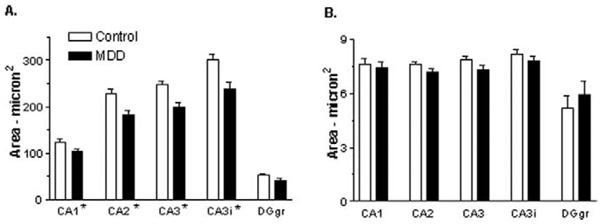
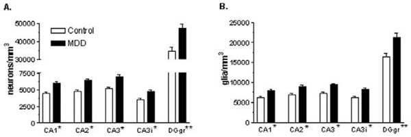
Results: In MDD, cryostat-cut hippocampal sections shrink in depth a significant 18% greater amount than in control subjects. The density of granule cells and glia in the dentate gyrus and pyramidal neurons and glia in all cornv ammonis (CA)/hippocampal subfields is significantly increased by 30%-35% in MDD. The average soma size of pyramidal neurons is significantly decreased in MDD.

Conclusion: In MDD, the packing density of glia, pyramidal neurons, and granule cell neurons is significantly increased in all hippocampal subfields and the dentate gyrus, and pyramidal neuron soma size is significantly decreased as well. It is suggested that a significant reduction in neuropil in MDD may account for decreased hippocampal volume detected by neuroimaging. In addition, differential shrinkage of frozen sections of the hippocampus suggests differential water content in hippocampus in MDD.


Figures

Figure 1
Figure 1
Brightfield photomicrographs of coronal sections of the postmortem human hippocampal formation. (A) Cresyl violet–stained section from a 70-year-old male control subject (postmortem interval = 20 hours) and (B) an adjacent section processed by Timm staining. Note the intensely stained granule cell layer of the dentate gyrus (DGgr) in (A) and (B), and the clear demarcation in (B) between hippocampal subfields CA2 and CA3 afforded by the Timm staining. A dashed line identifies the border between CA2 and CA3, and the second dashed line shows the border between CA3 inserted within the dentate gyrus (CA3i) and CA3 external to the dentate gyrus. (C) Cresyl violet–stained section from a depressed 77-year-old man (postmortem interval = 26 hours) and (D) an adjacent section processed by Timm staining. Pyramidal neurons and glial nuclei of CA3 are highlighted (E, Control; F, MDD) with large black arrows and white arrowheads, respectively. The scale bars in (A) and (E) are 750 µm and 25 µm, respectively.
Figure 2
Figure 2
Neuronal soma size (A) and glial nuclear size (B) in the hippocampus of control subjects and subjects with major depressive disorder (MDD). Pyramidal neurons were quantified in hippocampal fields CA1–CA3, and granule cells were quantified in the granule cell layer of the dentate gyrus (DGgr) of 21 control and 19 depressed subjects with the exception of 18 depressed subjects for CA1 and CA2. Values are least squares adjusted means ± SE. (A) There is a significant effect of diagnosis on pyramidal neuron soma size (*p = .0006) in all CA fields and a trend for an effect of diagnosis on granule cell soma size in the dentate gyrus (p = .0081). Pyramidal neuron soma size is decreased by 17%–21%, and granule cell soma size is decreased in the dentate gyrus by 22%. (B) Glial nuclear size was not significantly affected in MDD. CA3i refers to CA3 pyramidal neurons that are inserted within the dentate gyrus.
Figure 3
Figure 3
Neuronal (A) and glial (B) density in the hippocampus of control subjects and subjects with major depressive disorder (MDD). Pyramidal neurons were quantified in hippocampal fields CA1–CA3, and granule cells were quantified in the granule cell layer of the dentate gyrus (DGgr) of 21 control subjects and CA3 and dentate gyrus (DGgr) from 19 depressed subjects. Data in CA1 and CA2 are presented from 18 depressed subjects. Values are least squares adjusted means ± SE. (A) There is a significant effect of diagnosis on pyramidal neuron density in all CA subfields (*p < .0001) and granule cell density in the dentate gyrus (**p = .0004). Pyramidal neuron density is increased by 35%–36% in CA subfields, and granule cell density is increased in the dentate gyrus by 37%. (B) There is a significant effect of diagnosis on glial cell density in all CA pyramidal neuron subfields (*p < .0001) and glial cell density in the granule cell layer of the dentate gyrus (**p = .0007). Glial cell density is increased by 28%–31% in the CA pyramidal neuron subfields and glial cell density is increased in the granule cell layer of the dentate gyrus by 30%.

References

    1. Amaral DG, Insausti R. Hippocampal formation. In: Paxinos G, editor. The Human Nervous System. San Diego: Academic Press; 1990. pp. 711–755.
    1. Andersen BB, Gundersen HJ. Pronounced loss of cell nuclei and anisotropic deformation of thick sections. J Microsc. 1999;196:69–73. - PubMed
    1. Arnold SE, Franz BR, Gur RC, Gur RE, Shapiro RM, Moberg PJ, Trojanowski JQ. Smaller neuron size in schizophrenia in hippocampal subfields that mediate cortical-hippocampal interactions. Am J Psychiatry. 1995;152:738–748. - PubMed
    1. Benes FM, Kwok EW, Vincent SL, Todtenkopf MS. A reduction of nonpyramidal cells in sector CA2 of schizophrenics and manic depressives. Biol Psychiatry. 1998;44:88–97. - PubMed
    1. Bowley MP, Drevets WC, Ongur D, Price JL. Low glial numbers in the amygdala in major depressive disorder. Biol Psychiatry. 2002;52:404–412. - PubMed

Publication types

LinkOut - more resources