

Stage 0 macular holes: observations by optical coherence tomography
- PMID: 15522368
- PMCID: PMC1941774
- DOI: 10.1016/j.ophtha.2004.05.034
Stage 0 macular holes: observations by optical coherence tomography
Abstract
Objective: To introduce the concept of a stage 0 macular hole based on optical coherence tomographic observations of the vitreoretinal interface in fellow eyes of patients with unilateral idiopathic macular holes, and to evaluate the subsequent risk of progression to a full-thickness macular hole.
Design: Retrospective observational case series.
Participants: Ninety-four patients with a unilateral stage 2, 3, or 4 full-thickness macular hole.
Methods: The medical records of patients with a unilateral macular hole diagnosed between 1994 and 2000 at the New England Eye Center were reviewed.
Main outcome measure: Development of a full-thickness macular hole in the fellow eye on biomicroscopic fundoscopy or optical coherence tomography (OCT).
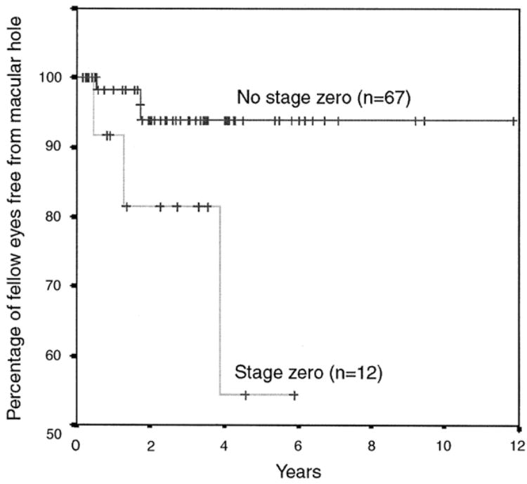
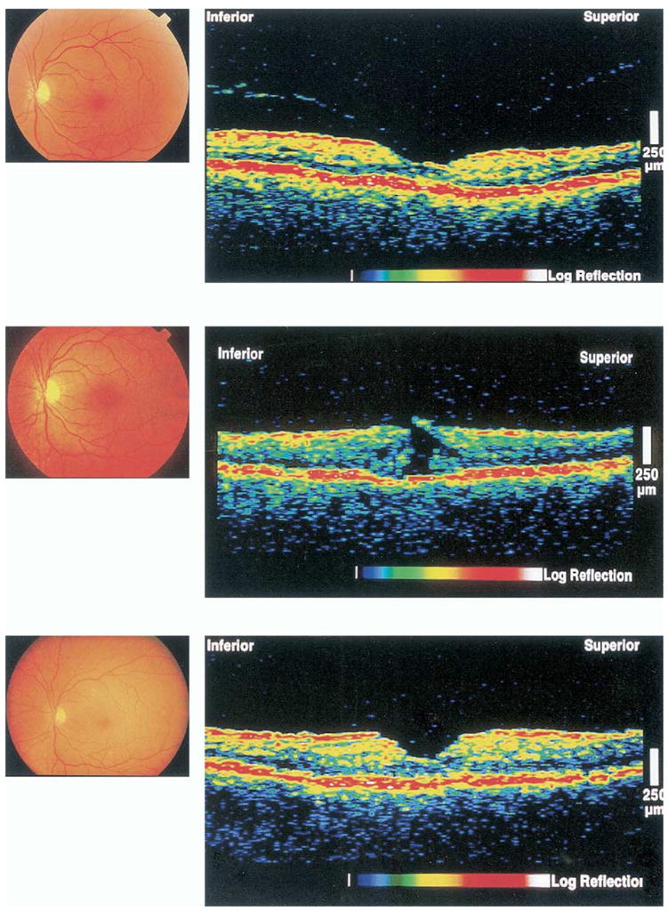
Results: In 27 (28.7%) of 94 clinically normal fellow eyes, OCT detected an abnormality of the vitreoretinal interface but normal foveal anatomy. The vitreoretinal abnormalities were further subclassified into severe (4 eyes), moderate (8 eyes), and mild (15 eyes) based on the intensity and morphology of the OCT signal. One of the 4 (25%) severe cases progressed to a full-thickness macular hole, 4 of the 8 (50%) moderate cases became full-thickness macular holes, and no (0%) mild cases progressed to a full-thickness macular hole. Severe and moderate eyes seemed to share characteristic features on OCT that increased their risk of macular hole development (stage 0 macular hole). The macular hole-free survival at 48 months was 94% for stage 0-negative patients, versus 54% for stage 0-positive patients. Univariate analysis revealed that the presence of a stage 0 macular hole was significantly associated with an almost 6-fold increase in the risk of macular hole formation (relative risk: 5.8, 95% confidence interval: 1.16-28.61, P = 0.03).
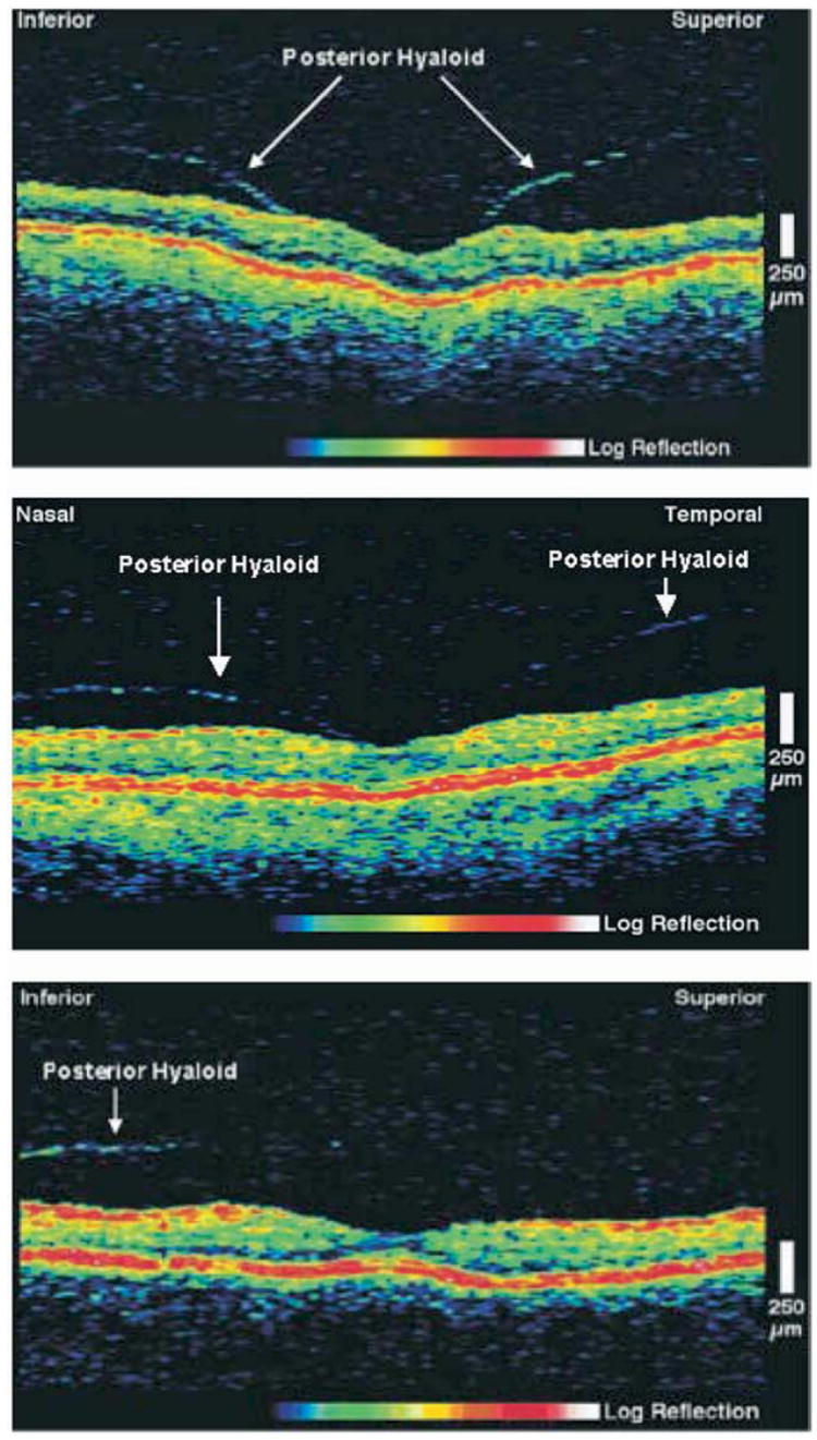
Conclusions: A stage 0 macular hole has a normal biomicroscopic appearance clinically, but has salient features on OCT as a result of oblique vitreous traction. Optical coherence tomographic findings consist of a normal foveal contour and normal retinal thickness and must include the presence of a preretinal, minimally reflective, thin band inserting obliquely on at least one side of the fovea. The presence of a stage 0 macular hole in the fellow eye is a significant risk factor for the development of a second macular hole.
Figures
Comment in
-
Stage 0 macular holes.Ophthalmology. 2005 Aug;112(8):1478. doi: 10.1016/j.ophtha.2005.01.009. Ophthalmology. 2005. PMID: 16061097 No abstract available.
References
-
- Kelly NE, Wendel RT. Vitreous surgery for idiopathic macular holes: results of a pilot study. Arch Ophthalmol. 1991;109:654–9. - PubMed
-
- Gass JDM. Idiopathic senile macular hole: its early stages and pathogenesis. Arch Ophthalmol. 1988;106:629–39. - PubMed
-
- Johnson RN, Gass JD. Idiopathic macular holes. Observations, stages of formation, and implications for surgical intervention. Ophthalmology. 1988;95:917–24. - PubMed
-
- Gass JD. Reappraisal of biomicroscopic classification of stages of development of a macular hole. Am J Ophthalmol. 1995;119:752–9. - PubMed
Publication types
MeSH terms
Grants and funding
LinkOut - more resources
Full Text Sources
