. 2004 Nov 16;101(46):16345-50.
doi: 10.1073/pnas.0407355101. Epub 2004 Nov 2.

Tissue plasminogen activator promotes the effects of corticotropin-releasing factor on the amygdala and anxiety-like behavior

Affiliations

Tissue plasminogen activator promotes the effects of corticotropin-releasing factor on the amygdala and anxiety-like behavior

Tomasz Matys et al. Proc Natl Acad Sci U S A. .

Abstract

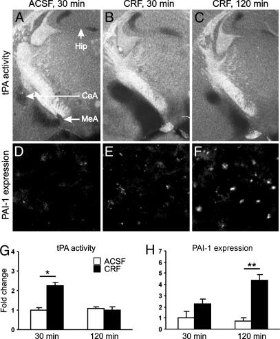
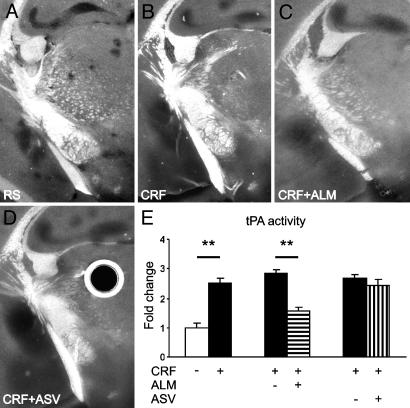
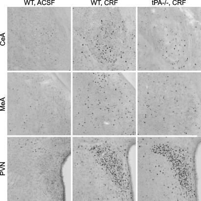
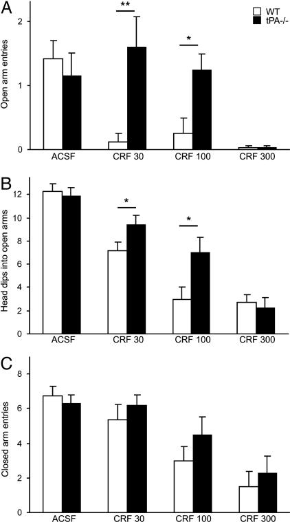
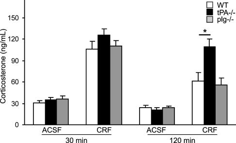
Stress-induced plasticity in the brain requires a precisely orchestrated sequence of cellular events involving novel as well as well known mediators. We have previously demonstrated that tissue plasminogen activator (tPA) in the amygdala promotes stress-induced synaptic plasticity and anxiety-like behavior. Here, we show that tPA activity in the amygdala is up-regulated by a major stress neuromodulator, corticotropin-releasing factor (CRF), acting on CRF type-1 receptors. Compared with WT, tPA-deficient mice responded to CRF treatment with attenuated expression of c-fos (an indicator of neuronal activation) in the central and medial amygdala but had normal c-fos responses in paraventricular nuclei. They exhibited reduced anxiety-like behavior to CRF but had a sustained corticosterone response after CRF administration. This effect of tPA deficiency was not mediated by plasminogen, because plasminogen-deficient mice demonstrated normal behavioral and hormonal changes to CRF. These studies establish tPA as an important mediator of cellular, behavioral, and hormonal responses to CRF.


Figures

Fig. 1.
Fig. 1.
CRF increases tPA activity in the amygdala in vivo. Thirty minutes after CRF injection, tPA activity in the amygdala (dark lytic areas in B) was significantly up-regulated in comparison with ACSF-injected control mice (A) and returned to basal values 90 min later (C). (D-F) Immunohistochemistry performed on adjacent brain sections showed increased PAI-1 expression in the same region. (G and H) Quantification of changes shown in A-C and D-F, respectively (n = 3-5 mice per group). CeA, central amygdala; MeA, medial amygdala; Hip, hippocampal mossy fiber pathway. The labels “30 min” and “120 min” indicate the time after injection. *, P < 0.05; **, P < 0.01.
Fig. 2.
Fig. 2.
CRF increases tPA activity in the amygdala in brain slices by means of the CRF-R1 receptor. CRF caused a significant up-regulation of tPA in the central and medial amygdala in comparison with the slice perfused with RS only (dark lytic areas in B vs. A). This up-regulation was inhibited by the CRF-R1 antagonist, antalarmin (ALM, C), but not the CRF-R2 antagonist Antisauvagine 30 (ASV, D). Slices perfused with CRF with addition of ALM or ASV vehicle (corresponding to C and D) are not shown. Round dark area with bright halo in D is an artifact caused by an air bubble in the overlay gel. (E) Quantification of changes shown in A-D (n = 5 slices per group) in relation to the mean area of lysis in slices perfused with RS only. **, P < 0.05.
Fig. 3.
Fig. 3.
tPA is necessary for neuronal activation in the central and medial amygdala. Examples of c-fos immunoreactive cells (visible as black dots) in the central (CeA) and medial (MeA) amygdala and PVN of WT and tPA-/- mice. For quantification and statistical analysis, see Table 1.
Fig. 4.
Fig. 4.
tPA-/- mice show a lower level of CRF-induced anxiety in the elevated plus-maze. (A) Injection of CRF strongly inhibited the exploration of open arms by WT animals, whereas tPA-/- mice failed to explore the open arms only at the highest dose of CRF. (B) tPA-/- animals more often dipped their heads into open arms. (C) Overall level of activity measured by closed arms entries was similar in both genotypes (n = 4-13 mice per group) *, P < 0.05; **, P < 0.01. CRF 30, CRF 100, and CRF 300 indicate dose of CRF in ng per animal.
Fig. 5.
Fig. 5.
tPA-/- mice show normal up-regulation of corticosterone in response to CRF but sustained elevation of corticosterone level during recovery. Shown is corticosterone concentration in WT, tPA-/-, and plg-/- mice (n = 4-5 per group) 30 or 120 min after CRF or ACSF injection. *, P < 0.05.

References

    1. Davis, M. (1992) Annu. Rev. Neurosci. 15 353-375. - PubMed
    1. Herman, J. P. & Cullinan, W. E. (1997) Trends Neurosci. 20 78-84. - PubMed
    1. Rogan, M. T. & LeDoux, J. E. (1996) Cell 85 469-475. - PubMed
    1. Owens, M. J. & Nemeroff, C. B. (1991) Pharmacol. Rev. 43 425-473. - PubMed
    1. Vale, W., Spiess, J., Rivier, C. & Rivier, J. (1981) Science 213 1394-1397. - PubMed

Publication types

MeSH terms

Substances