. 2004 Dec;2(12):e380.
doi: 10.1371/journal.pbio.0020380. Epub 2004 Nov 2.

Components of coated vesicles and nuclear pore complexes share a common molecular architecture

Affiliations

Components of coated vesicles and nuclear pore complexes share a common molecular architecture

Damien Devos et al. PLoS Biol. 2004 Dec.

Erratum in

  • PLoS Biol. 2005 Feb;3(2):e80

Abstract

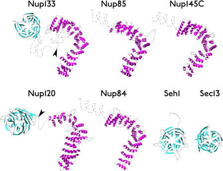
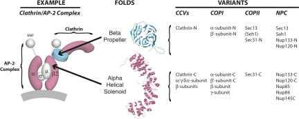
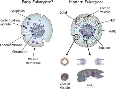
Numerous features distinguish prokaryotes from eukaryotes, chief among which are the distinctive internal membrane systems of eukaryotic cells. These membrane systems form elaborate compartments and vesicular trafficking pathways, and sequester the chromatin within the nuclear envelope. The nuclear pore complex is the portal that specifically mediates macromolecular trafficking across the nuclear envelope. Although it is generally understood that these internal membrane systems evolved from specialized invaginations of the prokaryotic plasma membrane, it is not clear how the nuclear pore complex could have evolved from organisms with no analogous transport system. Here we use computational and biochemical methods to perform a structural analysis of the seven proteins comprising the yNup84/vNup107-160 subcomplex, a core building block of the nuclear pore complex. Our analysis indicates that all seven proteins contain either a beta-propeller fold, an alpha-solenoid fold, or a distinctive arrangement of both, revealing close similarities between the structures comprising the yNup84/vNup107-160 subcomplex and those comprising the major types of vesicle coating complexes that maintain vesicular trafficking pathways. These similarities suggest a common evolutionary origin for nuclear pore complexes and coated vesicles in an early membrane-curving module that led to the formation of the internal membrane systems in modern eukaryotes.


Conflict of interest statement

The authors have declared that no conflicts of interest exist.

Figures

Figure 1
Figure 1. Ribbon Representation of Nup Models
β-sheets (β-propellers) are colored cyan and α-helices (α-solenoids) are colored magenta. Gray dashed lines indicate regions that were not modeled. Arrowheads indicate the positions of high proteolytic susceptibility (see Figures 2 and 3).
Figure 2
Figure 2. Proteolytic Domain Map of the Yeast Nup84 Subcomplex Proteins
Immunoblots of limited proteolysis digests for Protein A-tagged versions of each of the seven nups in the yNup84 subcomplex. Each protein is detected via its carboxyl-terminal tag; thus, all the fragments visualized are amino-terminal truncations (except for the full length proteins, which are indicated by arrowheads). The fragments of the Asp-N and Lys-C protease digests depicted in Figure 2 are labeled with letters (A, B, C…) that correspond to those in Table 2, and the terminal Protein A fragments are labeled with an X (the Protein A tag is resistant to proteolysis). The sizes of marker proteins are indicated in kilodaltons (kDa) to the right of the gel.
Figure 3
Figure 3. Predicted Secondary Structure Maps of the Nup84 Subcomplex Proteins
Thin horizontal lines represent the primary sequence of each protein; secondary structure predictions are shown as columns above each line for β-strands (β-propellers; cyan) and α-helices (α-solenoids; magenta). The height of the columns is proportional to the confidence of the secondary structure prediction (McGuffin et al. 2000). The modeled regions are indicated above each sequence by horizontal dark bars, corresponding to the models in Figure 1. Proteolytic cleavage sites are identified by small, medium, and large arrows for weak, medium, and strong susceptibility sites, respectively. Where necessary, uncertainties in the precise cleavage positions are indicated above the arrows by horizontal bars.
Figure 4
Figure 4. The Nup84 Complex and Coated Vesicles Share a Common Architecture
A diagram showing the organization of the clathrin/AP-2 coated vesicle complex is shown at left; the positions of clathrin and the adaptin AP-2 large subunits (α, β2 plus “ear” domains) and small subunits (σ, μ) are indicated. β-propeller regions are colored cyan, α-solenoid regions are colored magenta, and sample ribbon models for each fold are shown in the center. The variants of each fold that are found as domains in major components of the three kinds of vesicle-coating complexes and the yNup84 subcomplex are listed on the right. The -N and -C indicate amino-terminal and carboxyl-terminal domains, respectively. The classification of these domains is based on X-ray crystallography data (clathrin, α-adaptin, β2-adaptin [PDB codes 1gw5, 1bpo, 1b89 (ter Haar et al. 1998; Collins et al. 2002)]), by the detailed homology modeling presented here (yNup84 complex proteins; ySec13 also in Saxena et al. [1996]), or by sequence homology or unpublished secondary structure prediction and preliminary analyses (COPI I (sec31) complex proteins [Schledzewski et al. 1999], Sec31).
Figure 5
Figure 5. Proposed Model for the Evolution of Coated Vesicles and Nuclear Pore Complexes
Early eukaryotes (left) acquired a membrane-curving protein module (purple) that allowed them to mold their plasma membrane into internal compartments and structures. Modern eukaryotes have diversified this membrane-curving module into many specialized functions (right), such as endocytosis (orange), ER and Golgi transport (green and brown), and NPC formation (blue). This module (pink) has been retained in both NPCs (right bottom) and coated vesicles (left bottom), as it is needed to stabilize curved membranes in both cases.

References

    1. Amos LA, van den Ent F, Lowe J. Structural/functional homology between the bacterial and eukaryotic cytoskeletons. Curr Opin Cell Biol. 2004;16:24–31. - PubMed
    1. Andrade MA, Perez-Iratxeta C, Ponting CP. Protein repeats: Structures, functions, and evolution. J Struct Biol. 2001a;134:117–131. - PubMed
    1. Andrade MA, Petosa C, O'Donoghue SI, Muller CW, Bork P. Comparison of ARM and HEAT protein repeats. J Mol Biol. 2001b;309:1–18. - PubMed
    1. Apweiler R, Bairoch A, Wu CH, Barker WC, Boeckmann B, et al. UniProt: The Universal Protein knowledgebase. Nucleic Acids Res. 2004;32:D115–D119. Database issue. - PMC - PubMed
    1. Bednenko J, Cingolani G, Gerace L. Nucleocytoplasmic transport: Navigating the channel. Traffic. 2003;4:127–135. - PubMed

Publication types

MeSH terms

Substances