. 2005;15(2):273-80.
doi: 10.1002/hipo.20057.

Failure to acquire new semantic knowledge in patients with large medial temporal lobe lesions

Affiliations

Failure to acquire new semantic knowledge in patients with large medial temporal lobe lesions

Peter J Bayley et al. Hippocampus. 2005.

Abstract

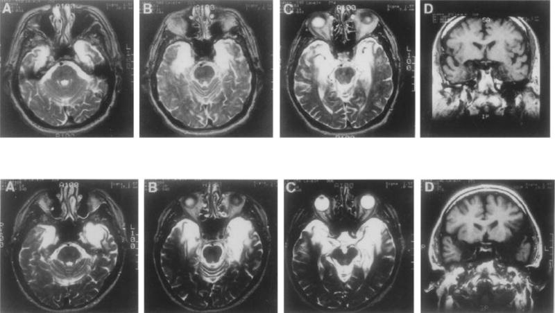
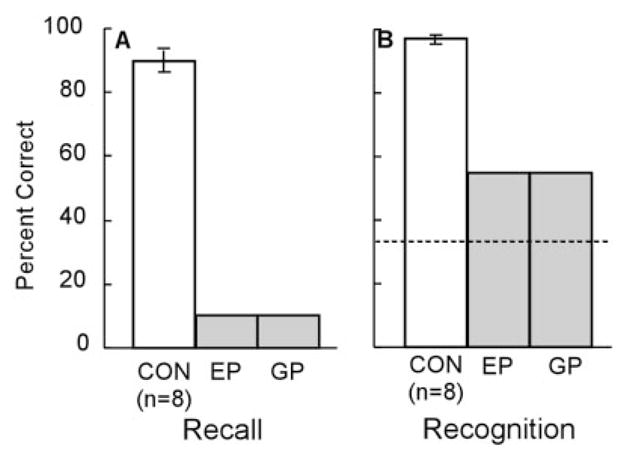
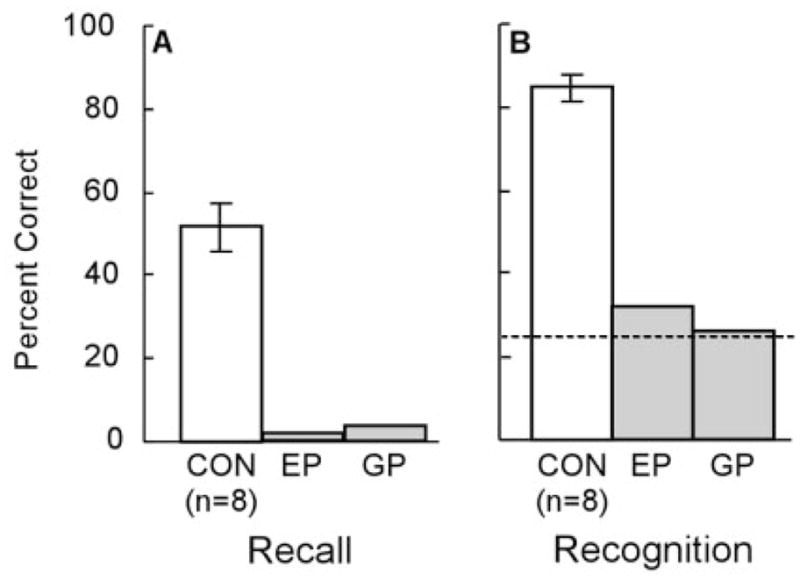
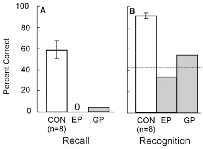
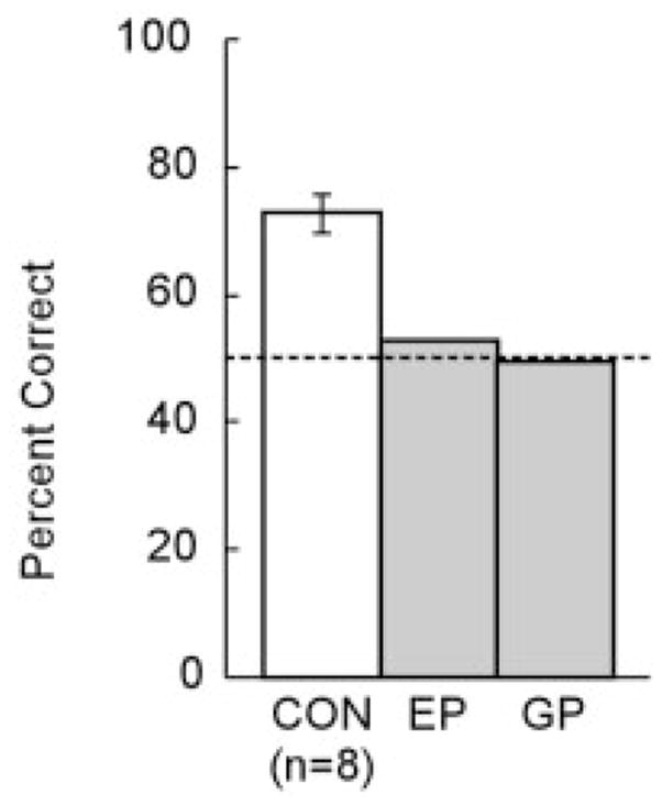
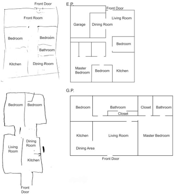
We examined new semantic learning in two profoundly amnesic patients (E.P. and G.P.) whose lesions involve virtually the entire medial temporal lobe (MTL) bilaterally. The patients were given five tests of semantic knowledge for information that could only have been acquired after the onset of their amnesia in 1992 and 1987, respectively. Age-matched and education-matched controls (n = 8) were also tested. On tests of recall, E.P. and G.P. each scored 10% correct on a test of 20 easy factual questions (controls = 90%), 2% and 4% correct on 55 questions about news events (controls = 85%), and 0% and 4% correct on a test of 24 famous faces. On three tests of recognition memory for this same material, the patients scored at chance levels. Similarly, the patients were unable to judge whether persons who had been famous for many decades were still living or had died during the past 10 years (E.P. = 53%; G.P. = 50%; controls = 73%; chance = 50%). Lastly, neither patient E.P. nor patient G.P. could draw an accurate floor plan of his current residence, despite having lived there for 10 years and 1 year, respectively. The results demonstrate that the capacity for new semantic learning can be absent, or nearly absent, when there is virtually complete damage to the MTL bilaterally. Accordingly, the results raise the possibility that the acquisition of conscious (declarative) knowledge about the world cannot be supported by structures outside the MTL, even with extended exposure. Published 2004 Wiley-Liss, Inc.


Figures

FIGURE 1
FIGURE 1
Magnetic resonance images showing the extent of temporal lobe damage in patients E.P. (top row) and G.P. (bottom row). A–C in each row are T2-weighted axial images through the temporal lobe. The images are continuous 5-mm sections (with 2.5-mm gaps) and are arranged from ventral (A) to dorsal (C). Damaged tissue is indicated by bright signal. D in each row is a coronal T1-weighted image at the level of the amygdala. Damaged tissue is indicated by dark signal. See text for description of the lesions.
FIGURE 2
FIGURE 2
Twenty easy facts. Performance of patients E.P., G.P., and controls (CON) in response to 20 questions about information that could only have been acquired after 1988. A: Percentage correct recall. B: Percentage correct responses on a three-alternative, forced-choice recognition test. Brackets show standard error of the mean, and the dashed line indicates chance performance (33%).
FIGURE 3
FIGURE 3
News events. Performance of patients E.P., G.P., and controls (CON) in response to 55 questions about news events that occurred from 1990 to 2000. A: Percentage correct recall. B: Percentage correct responses on a four-alternative, forced-choice recognition test. Brackets show standard error of the mean, and the dashed line indicates chance performance (25%).
FIGURE 4
FIGURE 4
Famous faces. Performance of patients E.P., G.P., and controls (CON) in response to photographs of 24 persons who had become famous during the 1990s. A: Percentage correct recall of the name of the person. B: Percentage correct responses for recognition (both three-alternative, forced-choice recognition and yes/no recognition). Brackets show standard error of the mean, and the dashed line indicates chance performance (42%).
FIGURE 5
FIGURE 5
Living or nonliving famous persons. Patients E.P., G.P., and controls (CON) were first asked to identify famous persons from among 126 nonfamous names and 126 persons who had become famous before 1970. For each person correctly identified as famous, participants were then asked whether that person was still living (44% of the famous persons were still living, and 56% had died between 1990 and 2001). Brackets show standard error of the mean, and the dashed line indicates chance performance (50%).
FIGURE 6
FIGURE 6
Home floor plan. Patients E.P. (top) and G.P. (bottom) were asked to draw a floor plan of their current home from memory. Both patients had moved to their current home after the onset of their amnesia. An accurate floor plan is shown alongside each patient’s drawing.

References

    1. Amaral DG, Insausti R. Hippocampal formation. In: Paxinos G, editor. The human nervous system. San Diego, CA: Academic Press; 1990.
    1. Bayley PJ, Hopkins RO, Squire LR. Successful recollection of remote autobiographical memories by amnesic patients with medial temporal lobe lesions. Neuron. 2003;38:135–144. - PubMed
    1. Bayley PJ, Squire LR. Medial temporal lobe amnesia: gradual acquisition of factual information by nondeclarative memory. J Neurosci. 2002;22:5741–5748. - PMC - PubMed
    1. Chan D, Fox N, Scahill R, Crum W, Whitwell J, Leschziner M, Rossor A, Steven J, Cipolotti L, Rossor M. Patterns of temporal lobe atrophy in semantic dementia and Alzheimer’s disease. Ann Neurol. 2001;49:433–442. - PubMed
    1. Corkin S. What’s new with the amnesic patient H.M.? Nat Rev Neurosci. 2002;3:153–160. - PubMed

Publication types