. 2005 Jan;16(1):248-59.
doi: 10.1091/mbc.e04-09-0807. Epub 2004 Nov 3.

Formation of membrane-bound ring complexes by prohibitins in mitochondria

Affiliations

Formation of membrane-bound ring complexes by prohibitins in mitochondria

Takashi Tatsuta et al. Mol Biol Cell. 2005 Jan.

Abstract

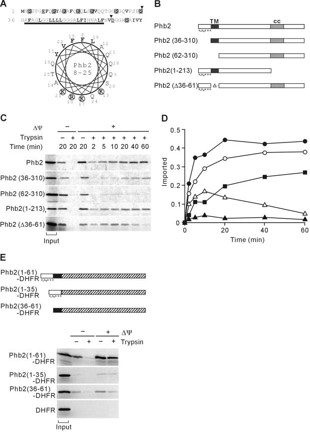
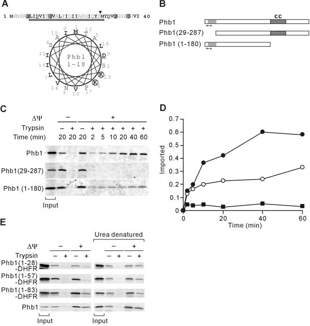
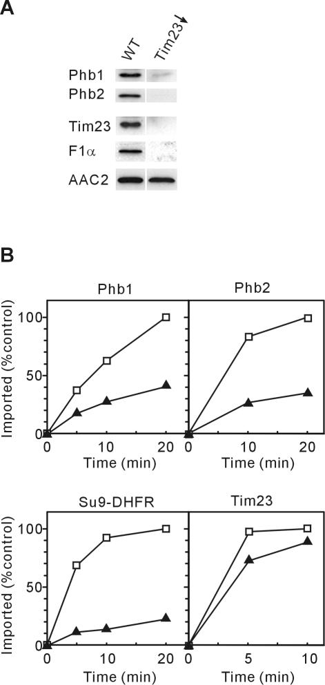
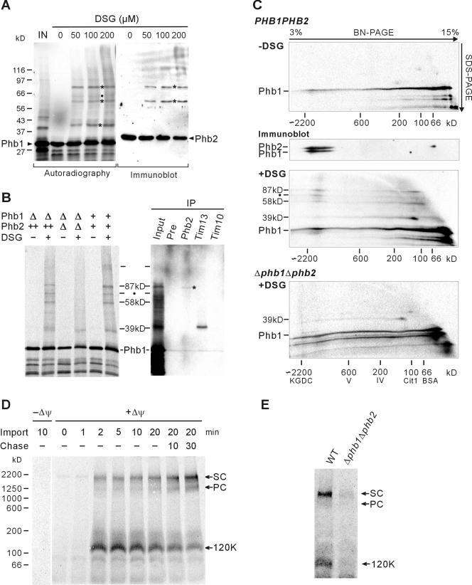
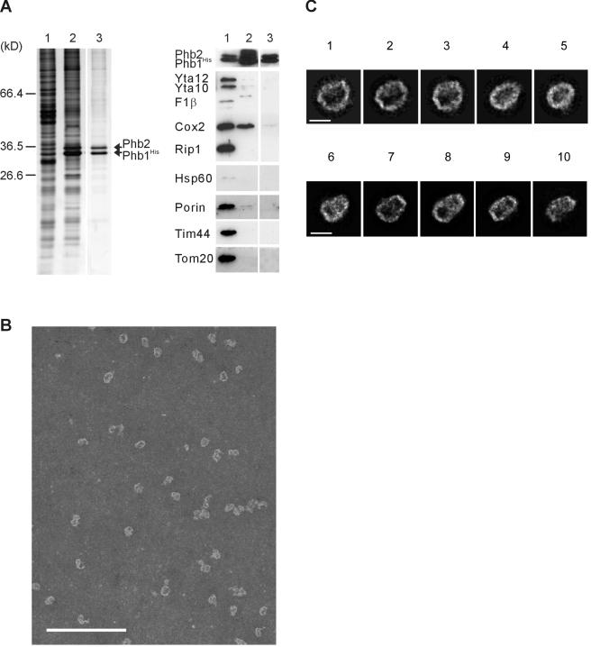
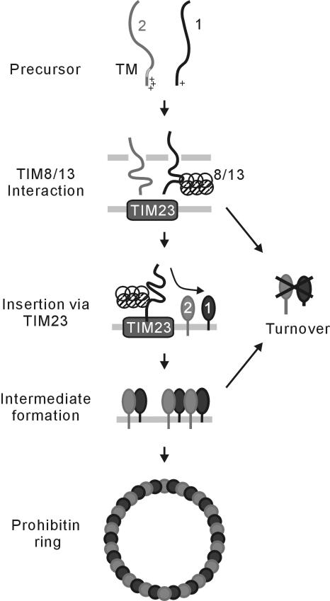
Prohibitins comprise a remarkably conserved protein family in eukaryotic cells with proposed functions in cell cycle progression, senescence, apoptosis, and the regulation of mitochondrial activities. Two prohibitin homologues, Phb1 and Phb2, assemble into a high molecular weight complex of approximately 1.2 MDa in the mitochondrial inner membrane, but a nuclear localization of Phb1 and Phb2 also has been reported. Here, we have analyzed the biogenesis and structure of the prohibitin complex in Saccharomyces cerevisiae. Both Phb1 and Phb2 subunits are targeted to mitochondria by unconventional noncleavable targeting sequences at their amino terminal end. Membrane insertion involves binding of newly imported Phb1 to Tim8/13 complexes in the intermembrane space and is mediated by the TIM23-translocase. Assembly occurs via intermediate-sized complexes of approximately 120 kDa containing both Phb1 and Phb2. Conserved carboxy-terminal coiled-coil regions in both subunits mediate the formation of large assemblies in the inner membrane. Single particle electron microscopy of purified prohibitin complexes identifies diverse ring-shaped structures with outer dimensions of approximately 270 x 200 angstroms. Implications of these findings for proposed cellular activities of prohibitins are discussed.


Figures

Figure 1.
Figure 1.
Targeting of Phb2 to mitochondria by a noncleavable, bipartite presequence at the N terminus. (A) N-terminal region of Phb2. Amino acid residues 1-70 of S. cerevisiae Phb2 and a helical wheel representation of amino acid residues 8-25 is shown. Charged and hydrophobic residues are highlighted. The bar indicates the predicted transmembrane region of Phb2. The position of N-terminal truncations of Phb2 is marked by arrowheads. (B) Schematic representation of Phb2 variants. TM, predicted transmembrane region; cc, putative coiled-coil region. Charged residues within the N-terminal targeting sequence are indicated. (C) Mitochondrial import of Phb2 and its variants. 35S-labeled precursor proteins were incubated with isolated mitochondria at 16°C for the indicated time in the presence (+Δψ) or absence (-Δψ) of a membrane potential. Nonimported proteins were digested with trypsin when indicated. The quantification of the import kinetics of Phb2 (•), Phb2(36-310) (▪), Phb2(62-310) (▴), Phb2(1-213) (○), Phb2(Δ36-61) (Δ) is shown in D. Total radioactivity incubated with mitochondria is shown as “input” in C and was set to 1 in D. (E) Top, schematic representation of Phb2-DHFR hybrid proteins. Shaded boxes represent the DHFR moiety. Other symbols are as in B. Bottom, import of Phb2-DHFR hybrid proteins into mitochondria. 35S-labeled hybrid proteins were incubated with isolated mitochondria for 20 min at 25°C in the presence (+Δψ) or absence (-Δψ) of a membrane potential. Trypsin was added to digest nonimported proteins when indicated. Mitochondria were analyzed by SDS-PAGE and autoradiography. Signals corresponding to 20% of radiolabeled precursors incubated with mitochondria are shown (input).
Figure 2.
Figure 2.
Mitochondrial targeting of Phb1 by an unconventional N-terminal presequence. (A) N-terminal region of Phb1. Amino acid residues 1-40 of S. cerevisiae Phb1 and a helical wheel representation of amino acid residues 1-18 is shown and marked as in Figure 1A. The gray bar indicates a potential transmembrane region of Phb1 that is predicted with low scores by using the TMHMM program. (B) Schematic representation of Phb1 and derivatives. Symbols are as in Figure 1B. (C) Mitochondrial import of Phb1 and its variants. 35S-labeled precursor proteins were incubated with isolated mitochondria at 25°C for the indicated time in the presence (+Δψ) or absence (-Δψ) of a membrane potential. Nonimported proteins were digested with trypsin when indicated. The quantification of the import kinetics of Phb1 (•), Phb1(29-288) (▪), and Phb1(1-180) (○) is shown in D. Total radioactivity added to the import reaction is shown as “input” in C and was set to 1 in D. (E) Import of Phb1-DHFR hybrid proteins into mitochondria. 35S-labeled hybrid proteins were incubated with isolated mitochondria for 20 min at 25°C as in C. When indicated, hybrid proteins were precipitated with ammonium sulfate and urea-denatured before import. Signals corresponding to 20% of radiolabeled precursors incubated with mitochondria are shown (input).
Figure 3.
Figure 3.
Requirement of the TIM23-translocase for prohibitin biogenesis. (A) Depletion of Tim23 in vivo. Mitochondial membranes were prepared from Tim23(Gal10) cells that were shifted for 24 h to galactose-free medium (Tim23↓). Samples were analyzed by SDS-PAGE and immunodecoration. YPH499 cells were analyzed equally for a control (WT). (B) Import of prohibitins into Tim23-deficient mitochondria. 35S-labeled precursor proteins of Phb1, Phb2, Su9(1-69)-DHFR, and Tim23 were imported into mitochondria isolated from wild-type (mb2) (□) or Tim23(fs) (▴) cells. Protease-resistant, imported protein was quantified by phosphorimaging.
Figure 4.
Figure 4.
Intermediate-sized prohibitin complexes containing Phb1 and Tim13 or Phb1 and Phb2. (A) Chemical cross-linking of newly imported Phb1. After import of 35S-labeled Phb1 and trypsin digestion, mitochondria were subjected to chemical cross-linking by using DSG at the concentrations indicated. Mitochondrial proteins were analyzed by SDS-PAGE and autoradiography (left) or immunodecoration with Phb2-specific antiserum (right). Major cross-link species are indicated by asterisks. A 63-kDa cross-link adduct, which was detected occasionally, is marked with a black dot. The signal corresponding to 50% of radiolabeled precursors incubated with mitochondria is shown (IN). (B) Chemical cross-linking of newly imported Phb1 in mitochondria isolated from wild-type (+/+), Δphb1Δphb2 (Δ/Δ), or Δphb1Δphb2 cells overexpressing Phb2 (-/++). After chemical cross-linking with DSG (150 μM), mitochondria were solubilized and subjected to immunoprecipitation (IP) (right) by using preimmune serum (Pre), Phb2-, Tim13-, and Tim10-specific antiserum. The Phb2-specific antiserum precipitated native Phb2 only very inefficiently (∼1%). A weak cross-link adduct coimmunoprecipitated with Phb2-specific antisera is indicated by an asterisk. (C) BN/SDS-PAGE analysis of cross-link adducts containing newly imported Phb1. After import of Phb1 into mitochondria isolated from YTT163 cells (PHB1PHB2, top) or YGS507 cells (Δphb1Δphb2, bottom), mitochondria were subjected to chemical cross-linking with DSG when indicated. Proteins were solubilized with 0.5% DDM (protein/detergent ratio of 1/2.5) and analyzed by two-dimensional BN/SDS-PAGE. α-Ketoglutarate dehydrogenase complex (KGDC, ∼2.2 MDa), monomeric F1FO-ATP synthase (complex V, ∼600 kDa), monomeric COX complex (complex IV, ∼200 kDa), dimeric citrate synthase (Cit1, 100 kDa), and bovine serum albumin (bovine serum albumin, ∼66 kDa) were used for calibration. The endogenous prohibitin complex was detected by immunoblotting by using Phb1- and Phb2-specific antibodies (middle). (D) Transient accumulation of newly imported Phb1 in intermediate-sized complexes. 35S-labeled Phb1 was imported into YTT160 mitochondria for various times in the absence (-Δψ) or presence (+Δψ) of a membrane potential. After import samples were treated with trypsin to remove nonimported precursors and further incubated at 25°C when indicated (Chase). Membranes were solubilized with 1.0% digitonin (protein/detergent ratio of 1/5) and analyzed by BN-PAGE and autoradiography. The position of the supercomplex of prohibitin with m-AAA protease (2 MDa, SC), prohibitin complex (1.2 MDa, PC), and ∼120-kDa intermediate (120K) are indicated. Dimeric F1FO-ATP synthase (∼1,250 kDa) and the supercomplex composed of bc1 and COX complexes (III2IV2, ∼1000 kDa) were used as additional molecular mass markers. (E) Impaired formation of prohibitin complexes in Δphb1Δphb2 mitochondria. 35S-labeled Phb1 was imported for 20 min into mitochondria isolated from wild-type (WT) or YGS507 cells (Δphb1Δphb2). Samples were processed as in D.
Figure 5.
Figure 5.
Assembly of the prohibitin complex depends on C-terminal regions of Phb1 and Phb2. (A and B) Mitochondrial membranes were analyzed by SDS-PAGE and immunoblotting by using Phb1- and Phb2-specific, and as a loading control, Yme1-specific antisera in the following yeast strains. (A) Wild-type cells (WT), Δphb1 cells (Δ), cells expressing C-terminally truncated Phb1(1-180)(YTT216), or Phb1(1-283)(YTT214) from the endogenous promoter (Genomic) and Δphb1 cells expressing Phb1 or the truncated Phb1 variants under the control of a constitutive TPI promoter (pTT46, pTT51, pTT50). (B) Wild-type cells (WT), Δphb2 cells (Δ), cells expressing Phb2 (pTT44), C-terminally truncated Phb2(1-191)(YTT222), or Phb2(1-308)(YTT220) from the endogenous promoter (Genomic) and Δphb2 cells expressing the truncated Phb2 variants Phb2(1-216)(pTT55) or Phb2(1-303)(pTT54) under the control of a constitutive TPI promoter. (C) BN/SDS-PAGE of newly imported Phb1 and Phb1 (1-180). 35S-labeled Phb1 and Phb1(1-180) were imported for 20 min at 25°C into mitochondria isolated from YTT163 cells. Mitochondrial membranes were solubilized by digitonin and subsequently analyzed by BN/SDS-PAGE. PC, prohibitin complex; SC, supercomplex of prohibitins with m-AAA protease. (D) Radiolabeled Phb1(1-180) was imported into mitochondria isolated from wild-type (+/+), Δphb1Δphb2 (Δ/Δ), or Δphb1Δphb2 strains expressing Phb2 (-/++) as in C followed by chemical cross-linking with DSG. When indicated (IP), mitochondrial extracts were analyzed by immunoprecipitation by using Phb2- and Tim13-specific antiserum or preimmune serum (Pre). The lane showing the immunoprecipitate with Phb2-specific antibodies was exposed three times as long as the other lanes.
Figure 6.
Figure 6.
Single particle electron microscopic analysis of the prohibitin complex. (A) Purification of prohibitin complexes from Δphb1 mitochondria harboring overexpressed Phb1His and Phb2 by metal chelating chromatography and glycerol gradient centrifugation as described in Materials and Methods. Samples were analyzed by SDS-PAGE and silver staining (left) or immunodetection against mitochondrial proteins as indicated (right). Prohibitin-containing fractions are shown. Lane 1, mitochondrial membranes (2 μg [silver staining] or 10 μg [immunodetection]); lane 2, eluate of Ni-NTA chromatography (1/200 or 1/40 of total); lane 3, prohibitin-containing fraction after glycerol gradient centrifugation (corresponding to a glycerol concentration of 24-26%; 1/40 or 1/8 of total). (B) Representative section of a micrograph showing a glycerol gradient fraction enriched in prohibitin complexes stained with 1% phosphotungstate. Bar, 200 nm. (C) Class averages of typical end-on or side-on projection views from prohibitin complexes. The data set was split in two subsets according to either elliptical ring-like (32%) or more rectangular views (68%). Percentages of particles of corresponding average images 1-5 are given as follows: for subset I (top), set to 100% class 1 counts 9%, class 2 6%, class 3 16%, class 4 12%, and class 5 11% of particles; for subset II (bottom), set to 100%, class 6 counts 17%, class 7 17%, class 8 8%, class 9 13% and class 10 8% of particles. Bar, 200 Å.
Figure 7.
Figure 7.
Model for the biogenesis of the ring-shaped prohibitin complex in mitochondria. See Discussion for details. The average stoichiometry of assembly intermediates and the ring complex is speculative. 1, Phb1; 2, Phb2; 8/13, TIM8/13; TM, transmembrane region.

References

    1. Arlt, H., Tauer, R., Feldmann, H., Neupert, W., and Langer, T. (1996). The YTA10-12-complex, an AAA protease with chaperone-like activity in the inner membrane of mitochondria. Cell 85, 875-885. - PubMed
    1. Artal-Sanz, M., Tsang, W. Y., Willems, E. M., Grivell, L. A., Lemire, B. D., van der Spek, H., Nijtmans, L. G., and Sanz, M. A. (2003). The mitochondrial prohibitin complex is essential for embryonic viability and germline function in Caenorhabditis elegans. J. Biol. Chem. 278, 32091-32099. - PubMed
    1. Back, J. W., Sanz, M. A., De Jong, L., De Koning, L. J., Nijtmans, L. G., De Koster, C. G., Grivell, L. A., Van Der Spek, H., and Muijsers, A. O. (2002). A structure for the yeast prohibitin complex: structure prediction and evidence from chemical crosslinking and mass spectrometry. Protein Sci. 11, 2471-2478. - PMC - PubMed
    1. Berger, K. H., and Yaffe, M. P. (1998). Prohibitin family members interact genetically with mitochondrial inheritance components in Saccharomyces cerevisiae. Mol. Cell. Biol. 18, 4043-4052. - PMC - PubMed
    1. Birner, R., Nebauer, R., Schneiter, R., and Daum, G. (2003). Synthetic lethal interaction of the mitochondrial phosphatidylethanolamine biosynthetic machinery with the prohibitin complex of Saccharomyces cerevisiae. Mol. Biol. Cell 14, 370-383. - PMC - PubMed

Publication types

MeSH terms

Substances