. 2004 Dec;10(12):1957-66.
doi: 10.1261/rna.7135204. Epub 2004 Nov 3.

Human microRNAs are processed from capped, polyadenylated transcripts that can also function as mRNAs

Affiliations

Human microRNAs are processed from capped, polyadenylated transcripts that can also function as mRNAs

Xuezhong Cai et al. RNA. 2004 Dec.

Abstract

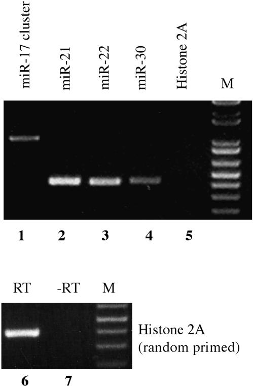
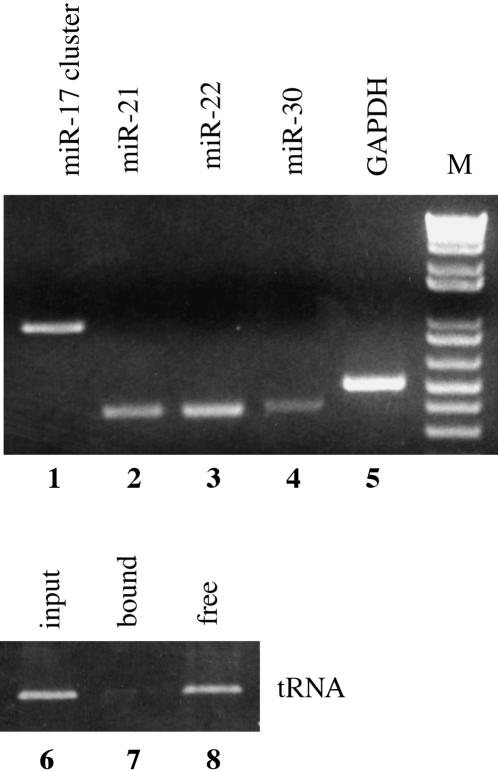
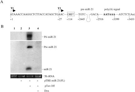
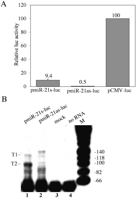
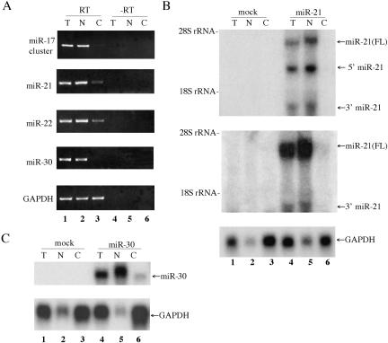
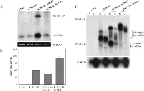
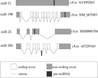
The factors regulating the expression of microRNAs (miRNAs), a ubiquitous family of approximately 22-nt noncoding regulatory RNAs, remain undefined. However, it is known that miRNAs are first transcribed as a largely unstructured precursor, termed a primary miRNA (pri-miRNA), which is sequentially processed in the nucleus, to give the approximately 65-nt pre-miRNA hairpin intermediate, and then in the cytoplasm, to give the mature miRNA. Here we have sought to identify the RNA polymerase responsible for miRNA transcription and to define the structure of a full-length human miRNA. We show that the pri-miRNA precursors for nine human miRNAs are both capped and polyadenylated and report the sequence of the full-length, approximately 3433-nt pri-miR-21 RNA. This pri-miR-21 gene sequence is flanked 5' by a promoter element able to transcribe heterologous mRNAs and 3' by a consensus polyadenylation sequence. Nuclear processing of pri-miRNAs was found to be efficient, thus largely preventing the nuclear export of full-length pri-miRNAs. Nevertheless, an intact miRNA stem-loop precursor located in the 3' UTR of a protein coding gene only moderately inhibited expression of the linked open reading frame, probably because the 3' truncated mRNA could still be exported and expressed. Together, these data show that human pri-miRNAs are not only structurally similar to mRNAs but can, in fact, function both as pri-miRNAs and mRNAs.


Figures

FIGURE 1.
FIGURE 1.
Polyadenylation of pri-miRNA transcripts. A HeLa cDNA preparation, obtained by oligo(dT) affinity purification of total HeLa cell RNA followed by oligo(dT) primed reverse transcription, was subjected to PCR using primers flanking the human miR-21, miR-22, or miR-30 miRNA stem–loop or the miR-17 miRNA cluster. Primers were designed to sit down ~170 bp 5′ and 3′ to the predicted miRNA stem–loops and were, therefore, expected to give rise to ~410-bp fragments for miR-21, miR-22, and miR-30, or a 1000-bp fragment for the miR-17 cluster. Histone H2A mRNA, which is not polyadenylated, served as a negative control. (Lanes 6,7) To confirm that the Histone 2A primers were functional, PCR was also performed using random primed HeLa cDNA. M, DNA size markers.
FIGURE 2.
FIGURE 2.
Pri-miRNA transcripts are capped. Capped RNAs were purified from total HeLa cell mRNA using beads loaded with a high-affinity mutant form of eIF4E, as previously described (Choi and Hagedorn 2003). The recovered capped RNAs were reverse transcribed using random primers and then subjected to PCR using the primers described in Figure 1 or using primers specific for human GAPDH mRNA or alanine tRNA. In lanes 68, the input, bound and free, supernatant alanine tRNAs were also analyzed.
FIGURE 3.
FIGURE 3.
Characterization of the full-length pri-miR-21 RNA transcript. (A) Schematic representation of the miR-21 gene. The 3′-end of the pri-miR-21 transcript was determined by RACE, while two approximate transcription start sites, indicated by T1 and T2, were mapped by RACE and by primer extension. The pri-miR-21 RNA poly(A) signal, the cleavage site used for polyadenylation and a possible 124-amino-acid ORF are also indicated. (B) The full-length pri-miR-21 RNA was placed under the transcriptional control of an inducible TRE-CMV promoter and transfected into 293T cells in the presence or absence of the pTet-Off activator plasmid. Dox was used to inhibit TRE-CMV-driven transcription. Expression of miR-21 was detected by Northern blot. 5S rRNA was utilized as a loading control.
FIGURE 4.
FIGURE 4.
Characterization of the pri-miR-21 promoter. (A) miR-21 promoter-driven luc expression. Plasmids pmiR-21s-luc, pmiR-21as-luc, and pCMV-luc were cotransfected into 293T cells along with a Renilla luciferase internal control plasmid. Induced luciferase activities were determined at 48 h after transfection and normalized to the Renilla luciferase internal control (average of three independent experiments). Data are presented relative to the firefly luciferase activity detected in cultures transfected with pCMV-luc, which was set at 100. (B) Primer extension analysis using a luciferase gene-specific primer and RNA recovered from 293T cells transfected with pmiR-21s-luc or pmiR-21as-luc or was mock transfected. Major (T2) and minor (T1) extension products observed in the pmiR-21s-luc transfected cells are indicated and are mapped to the underlying miR-21 promoter sequence in Figure 3A.
FIGURE 5.
FIGURE 5.
Primary miRNA transcripts are largely confined to the nucleus. (A) Distribution of endogenous pri-miRNAs. HeLa cell RNA was isolated from the total (T) cell, or from the nucleus (N) and cytoplasm (C), and was then subjected to reverse transcription using oligo(dT) primers. PCR was performed using the primers described in Figure 1. RNA that had not been subjected to reverse transcription served as a negative control. (B) Distribution of overex-pressed pri-miR21 RNA. 293T cells were mock transfected or were transfected with pTRE-miR-21(FL) in the presence of the pTet-Off activator plasmid. RNA was isolated at ~36 h after transfection and subjected to Northern analysis using nick-translated miR-21 specific probes, derived from nucleotides +2234 to +2648 (upper panel) or +2904 to +3283 (middle panel) of the predicted pri-miR-21 RNA, or using a probe specific for the endogenous GAPDH mRNA as a loading control (lower panel). (C) Similar to B, except that 293T cells were transfected with pTRE-miR-30, which encodes an artificial pri-miR-30 RNA. The probe used was derived from the 414-bp miR-30 DNA fragment present in pTRE-miR-30, which extends both 5′ and 3′ to the predicted pre-miRNA processing sites.
FIGURE 6.
FIGURE 6.
A pri-miRNA can function as an mRNA. (A) 293T cells were cotransfected with the indicated pTRE-based expression plasmid and with the pTet-Off activator plasmid. Pre-miR-30 and miR-30as expression was detected at 60 h after transfection by Northern blot. 5S rRNA was utilized as a loading control. (B) In parallel, 293T cells were transfected with the indicated pTRE-based expression plasmid together with pTet-Off and a Renilla luciferase expression plasmid. At 48 h after transfection, firefly and Renilla luciferase activities were measured. Data were normalized to the Renilla luciferase internal control and are presented relative to the activity detected in the pTRE-luc transfected culture, which was set at 100. Average of three experiments with standard deviation indicated. (C) Northern analysis using nuclear (N) or cytoplasmic (C) RNA fractions obtained from 293T cells transfected with the indicated pTRE-based expression plasmids. The probe used in the upper panel is specific to the firefly luciferase gene. The predicted full-length and 3′ truncated luc mRNAs expressed in each culture are indicated. The blot was stripped and reprobed with a probe specific for the endogenous GAPDH mRNA (lower panel).
FIGURE 7.
FIGURE 7.
Location of microRNA stem–loops in RNA transcripts. The figure shows the relative position of miRNA stem–loops in known human RNA transcripts. The miR-21 miRNA analyzed in this manuscript is transcribed as part of an unspliced, apparently noncoding RNA, while miR-22 is found in an exonic location in a spliced non-coding RNA. miRNAs located in protein coding genes are mostly found in introns, as shown here for miR-26b, although a location in the 3′ UTR of an mRNA, as shown here for miR-198 and the human gene FRP, is occasionally observed. The figure is not drawn to scale. The exon/intron structure of candidate pri-miRNAs was generated by aligning the sequence of putative pri-miRNAs with the corresponding genomic DNA sequence. The pri-miR-21 RNA shown was characterized in this manuscript. GenBank accession numbers of all the candidate pri-miRNA transcripts are shown.

References

    1. Aukerman, M.J. and Sakai, H. 2003. Regulation of flowering time and floral organ identity by a microRNA and its APETALA2-like target genes. The Plant Cell 15: 2730–2741. - PMC - PubMed
    1. Bartel, D.P. 2004. MicroRNAs: genomics, biogenesis, mechanism, and function. Cell 116: 281–297. - PubMed
    1. Bohnsack, M.T., Czaplinski, K., and Görlich, D. 2004. Exportin 5 is a RanGTP-dependent dsRNA-binding protein that mediates nuclear export pre-miRNAs. RNA 10: 185–191. - PMC - PubMed
    1. Bracht, J., Hunter, S., Eachus, R., Weeks, P., and Pasquinelli, A.E. 2004. Trans-splicing and polyadenylation of let-7 microRNA primary transcripts. RNA 10: 1586–1594. - PMC - PubMed
    1. Chen, C.-Z., Li, L., Lodish, H.F., and Bartel, D.P. 2004. MicroRNAs modulate hematopoietic lineage differentiation. Science 303: 83–86. - PubMed

Publication types

LinkOut - more resources