. 2004 Nov 3;24(44):9770-8.
doi: 10.1523/JNEUROSCI.3027-04.2004.

Electrical coupling among irregular-spiking GABAergic interneurons expressing cannabinoid receptors

Affiliations

Electrical coupling among irregular-spiking GABAergic interneurons expressing cannabinoid receptors

Mario Galarreta et al. J Neurosci. .

Abstract

Anatomical studies have shown that the G-protein-coupled cannabinoid receptor-1 (CB1) is selectively expressed in a subset of GABAergic interneurons. It has been proposed that these cells regulate rhythmic activity and play a key role mediating the cognitive actions of marijuana and endogenous cannabinoids. However, the physiology, anatomy, and synaptic connectivity of neocortical CB1-expressing interneurons remain poorly studied. We identified a population of CB1-expressing interneurons in layer II/III in mouse neocortical slices. These cells were multipolar or bitufted, had a widely extending axon, and exhibited a characteristic pattern of irregular spiking (IS) in response to current injection. CB1-expressing-IS (CB1-IS) cells were inhibitory, establishing GABAA receptor-mediated synapses onto pyramidal cells and other CB1-IS cells. Recently, electrical coupling among other classes of cortical interneurons has been shown to contribute to the generation of rhythmic synchronous activity in the neocortex. We therefore tested whether CB1-IS interneurons are interconnected via electrical synapses using paired recordings. We found that 90% (19 of 21 pairs) of simultaneously recorded pairs of CB1-IS cells were electrically coupled. The average coupling coefficient was 6%. Signaling through electrical synapses promoted coordinated firing among CB1-IS cells. Together, our results identify a population of electrically coupled CB1-IS GABAergic interneurons in the neocortex that share a unique morphology and a characteristic pattern of irregular spiking in response to current injection. The synaptic interactions of these cells may play an important role mediating the cognitive actions of cannabinoids and regulating coherent neocortical activity.


Figures
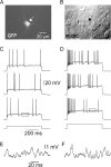
Figure 1.
Figure 1.
A population of layer II/III GABAergic interneurons is characterized by an irregular pattern of spiking. A, A multipolar EGFP-GAD65 fluorescing cell in a 17-d-old mouse. B, Same field visualized under infrared differential interference contrast (IR-DIC) video microscopy. The arrow points to the cell illustrated in A. C, Three examples of the firing pattern of the same neuron in response to a pulse of depolarizing current injection (200 pA, 600 msec). Note the characteristic irregular spiking. D, Firing pattern in response to a larger current injection (400 pA, 600 msec). E, F, Portions of the bottom traces in C and D (indicated by the boxes) are shown at an expanded scale to illustrate the oscillations of the membrane potential.
Figure 2.
Figure 2.
Firing properties in IS cells differ from those in FS cells. A, Irregular firing of an IS cell in layer II/III in response to a pulse of depolarizing current (400 pA, 600 msec). B, A typical discharge of high-frequency nonaccommodating action potentials in an FS cell in response to a near-threshold current injection (525 pA, 600 msec). C, D, Plot of the interspike intervals for the traces shown in A and B, respectively. E, Example of an IS cell action potential. F, Example of an FS cell action potential. The IS and FS spikes are superimposed in the inset in E. Note that the IS spike is wider than that of the FS cell. Also note that the AHP is smaller and slower in the IS cell than in the FS cell. Calibrations in B and F apply to A and E, respectively.
Figure 3.
Figure 3.
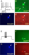
IS cells are immunoreactive to CB1 receptors. A, Characteristic irregular spiking of a layer II/III neuron. B, EGFP fluorescence of the cell whose response is shown in A (arrow). The arrowhead points to the axon of this neuron. The double arrowhead points to an unidentified EGFP-positive-CB1-negative cell. C, Photograph of the recorded cell filled with biocytin and revealed with streptavidin-Alexa Fluor 350. D, CB1 immunoreactivity of the cell shown in A-C (arrow). The immunoreactive axon is indicated by the arrowhead. Note the dense network of immunoreactive fibers in layer II/III. Scale bar, 25 μm (applies to all images). E, F, FS cells lack CB1 immunoreactivity. E, Pattern of firing of a representative FS cell filled with biocytin (G) during the recording (arrow). This cell was EGFP negative (F) and did not show immunoreactivity for CB1 receptors (H). The arrowheads in F and H point to a nonrecorded EGFP-positive cell immunoreactive for CB1 receptors.
Figure 4.
Figure 4.
CB1-IS cells are GABAergic. A, Morphological reconstruction of a pair consisting of a presynaptic CB1-IS cell (red) and a postsynaptic pyramidal cell (blue; CB1-IS → P). Dendrites, Thick lines; axons, thin lines. The insets show the firing of both cells in response to a pulse of depolarizing current. p, Piamater; L I, layer I; wm, white mater. B, Paired recording from the two cells illustrated in A. The postsynaptic membrane of the pyramidal cell was either depolarized (-48 mV) or hyperpolarized (-91 mV) ([Cl-]i = 6.3 mm). Traces are the average of 80 responses. C, Morphological reconstruction of two synaptically connected CB1-IS cells (CB1-IS → CB1-IS). The presynaptic cell is in red, and the postsynaptic cell is in blue. Dendrites, Thick lines; axons, thin lines. The insets show examples of their characteristic irregular spiking in response to pulses of current injection. D, Paired recording from the two CB1-IS cells illustrated in C. Two action potentials separated by 50 msec were generated in the presynaptic cell (top trace). Five individual postsynaptic responses recorded at -78 mV ([Cl-]i = 40 mm; middle traces). Note the variability in the amplitude of the postsynaptic responses (coefficient of variation, 0.81). Bottom trace, Average of 50 responses. The paired-pulse ratio (IPSP2/IPSP1) was 0.94. The decay of the average IPSP was fitted with an exponential function with a time constant of 21 msec.
Figure 5.
Figure 5.
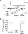
CB1 receptor activation blocks IPSPs produced by CB1-IS cell onto pyramidal cells. A, Paired recording between a presynaptic CB1-IS cell and a postsynaptic pyramidal neuron (Vm = -55 mV). A control IPSP (control) was abolished after 5 min of bath application of the cannabinoid receptor agonist WIN55,212-2 (1 μm; WIN). Addition of the CB1 receptor antagonist AM-251 (10 μm) reversed the effect of WIN55,212-2. Each trace is the average of 50-75 trials. B, Bar graph summarizing the results obtained in four experiments similar to the one shown in A. The amplitude of the response in the presence of WIN55,212-2 was significantly smaller than that of the IPSP recorded in control condition (p < 0.0001) and in the presence of AM-251 (p < 0.05).
Figure 6.
Figure 6.
CB1-IS cells are electrically coupled. A, Morphological reconstruction of a pair of electrically coupled CB1-IS cells (CB1-IS = CB1-IS). Dendrites, Thick lines; axons, thin lines. The insets show the characteristic irregular firing of both cells in response to a pulse of depolarizing current. p, Piamater; L I, layer I; wm, white mater. B, DAB staining of the CB1-IS cells shown in A. C, Paired recording of the cells illustrated in A and B. Left, The injection of depolarizing (+100 pA) or hyperpolarizing (-100 pA) current in cell 1 simultaneously affected the membrane voltage of the noninjected cell 2. The injection of current in cell 2 similarly affected the membrane potential of cell 1. The coupling coefficient was 10.2%. Traces are the average of 80-100 responses. D, Bi-directionality of electrical coupling. Plot showing the coupling coefficient when current is transmitted from cell 1 to cell 2 and from cell 2 to cell 1. Data from the same pair are connected by a line.
Figure 7.
Figure 7.
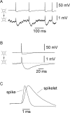
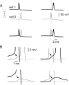
Spike transmission between electrically coupled CB1-IS cells. A, Paired recording from two electrically coupled CB1-IS cells. Spikes produced in one of the cells by sustained depolarizing current injection generated biphasic responses in the second cell (Vm = -44 and -74 mV for the top and bottom traces, respectively). The coupling coefficient was 9.8%. B, Average of 45 traces aligned at the peak of the spike. Data from the same pair as in A. Note the fast depolarizing component of the spikelet followed by a slow hyperpolarization, reflecting the presynaptic AHP. C, The presynaptic spike and the postsynaptic spikelet from B are shown scaled and superimposed to facilitate the comparison of their time course.
Figure 8.
Figure 8.
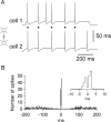
Electrical coupling coordinates the firing of CB1-IS cells. A, Simultaneous recording from a pair of CB1-IS cells depolarized to near threshold (average action potential frequencies were 7.6 and 7.3 Hz). Diamonds indicate spikes occurring in both cells within a window of ≤5 msec. The coupling coefficient was 9.8%. B, Cross-correlogram of the cells shown in A. The bin size is 1 msec. The probability of spikes is increased near 0 msec delay. The inset shows the central area of the cross-correlogram at an expanded time scale, with two peaks at ±1-2 msec.
Figure 9.
Figure 9.
Coherent firing among electrically coupled CB1-IS cells is mediated by spikelets. A, Paired recording from two electrically coupled CB1-IS cells. Two examples of coordinated firing are shown. On the left, the spiking in cell 1 (thick line) precedes the firing in cell 2 (thin line) by 1.6 msec. Both traces are superimposed in the bottom. On the right, the firing in cell 2 precedes the spike in cell 1 by 1.9 msec. B, Top panels show data from A at an expanded voltage scale. The arrows point to the spikelet. Bottom panels show data from examples in which the spikelet in the postsynaptic cell did not reach spike threshold.

References

    1. Bartos M, Vida I, Frotscher M, Meyer A, Monyer H, Geiger JR, Jonas P (2002) Fast synaptic inhibition promotes synchronized gamma oscillations in hippocampal interneuron networks. Proc Natl Acad Sci USA 99: 13222-13227. - PMC - PubMed
    1. Beierlein M, Gibson JR, Connors BW (2000) A network of electrically coupled interneurons drives synchronized inhibition in neocortex. Nat Neurosci 3: 904-910. - PubMed
    1. Blatow M, Rozov A, Katona I, Hormuzdi SG, Meyer AH, Whittington MA, Caputi A, Monyer H (2003) A novel network of multipolar bursting interneurons generates theta frequency oscillations in neocortex. Neuron 38: 805-817. - PubMed
    1. Bodor AL, Katona I, Mackie K, Hajos N, Freund TF (2003) Distinct types of interneurons are targeted by endocannabinoids in the rat soomatosensory cortex. Soc Neurosci Abstr 29: 578.13.
    1. Brager DH, Luther PW, Erdélyi F, Szabó G, Alger BE (2003) Regulation of exocytosis from single visualized GABAergic boutons in hippocampal slices. J Neurosci 23: 10475-10486. - PMC - PubMed

Publication types

LinkOut - more resources