. 2004 Nov 3;24(44):9811-25.
doi: 10.1523/JNEUROSCI.1532-04.2004.

Entorhinal cortex lesions disrupt the relational organization of memory in monkeys

Affiliations

Entorhinal cortex lesions disrupt the relational organization of memory in monkeys

Cindy A Buckmaster et al. J Neurosci. .

Abstract

Recent accounts suggest that the hippocampal system critically supports two central characteristics of episodic memory: the ability to establish and maintain representations for the salient relationships between experienced events (relational representation) and the capacity to flexibly manipulate memory (flexible memory expression). To test this proposal in monkeys, intact controls and subjects with bilateral aspiration lesions of the entorhinal cortex were trained postoperatively on two standard memory tasks, delayed nonmatchingto-sample (DNMS) and two-choice object discrimination (OD) learning, and three procedures intended to emphasize relational representation and flexible memory expression: a paired associate (PA) task, a transitive inference (TI) test of learning and memory for hierarchical stimulus relationships, and a spatial delayed recognition span (SDRS) procedure. The latter assessments each included critical "probe" tests that asked monkeys to evaluate the relationships among previously learned stimuli presented in novel combinations. Subjects with entorhinal cortex lesions scored as accurately as controls on all phases of DNMS and OD, procedures that can be solved on the basis of memory for individual stimuli. In contrast, experimental monkeys displayed deficits relative to controls on all phases of the PA, TI, and SDRS tasks that emphasized the flexible manipulation of memory for the relationships between familiar items. Together, the findings support the conclusion that the primate hippocampal system critically enables the relational organization of declarative memory.


Figures
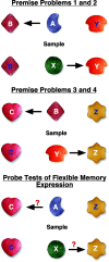
Figure 1.
Figure 1.
“Cookie” stimuli and schematic representation of problems for the relational series of the paired associate task. Arrows point to the correct choice for each problem. White and blue letters signify positive (sweet) and negative (bitter) discriminative stimuli, respectively.
Figure 2.
Figure 2.
Schematic representation of the instruction discriminations and probe tests of the transitive inference task. Unfilled letters signify stimuli associated with equivalent reinforcement (50%) over the course of training (see Materials and Methods for details).
Figure 3.
Figure 3.
Schematic representations of type-1 (left) and type-2 (right) probe trials in the spatial delayed recognition span task. For the critical final choice (bottom row), white circles signify locations baited previously in the same trial. +, Positive (sweet); -, negative (bitter).
Figure 4.
Figure 4.
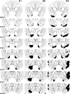
Representative coronal sections through the brain of an intact cynomolgus monkey (Szabo and Cowan, 1984) illustrating the intended lesion site. Sections are arranged from rostral (A20.0) to caudal (A10.8), and the entorhinal cortex is highlighted in gray.
Figure 5.
Figure 5.
Schematic representation of damage observed in monkeys E1-E3, illustrated across the same rostrocaudal levels as in Figure 4. L, Left hemisphere; R, right hemisphere.
Figure 6.
Figure 6.
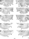
Photomicrographs through the left (shown as left) and right (shown as right) medial temporal lobes of experimental monkeys E1 (6.1), E2 (6.2), and E3 (6.3). A-E, Sections are arranged rostral to caudal. Arrowheads delimit the mediolateral extent of the lesion. See Results for detailed description of unintended damage in each case. A, Amygdala; 35, perirhinal area 35; 36, perirhinal area 36; H, hippocampus; rs, rhinal sulcus; amts, anterior medial temporal sulcus; sts, superior temporal sulcus (6.3). Scale bar, 1 mm.
Figure 6.
Figure 6.
Photomicrographs through the left (shown as left) and right (shown as right) medial temporal lobes of experimental monkeys E1 (6.1), E2 (6.2), and E3 (6.3). A-E, Sections are arranged rostral to caudal. Arrowheads delimit the mediolateral extent of the lesion. See Results for detailed description of unintended damage in each case. A, Amygdala; 35, perirhinal area 35; 36, perirhinal area 36; H, hippocampus; rs, rhinal sulcus; amts, anterior medial temporal sulcus; sts, superior temporal sulcus (6.3). Scale bar, 1 mm.
Figure 7.
Figure 7.
Mean trials to criterion on the delayed nonmatching-to-sample task with a 10 sec delay (left). Mean percentage correct across retention intervals of 15-600 sec (right). C, n = 6; E, n = 3.
Figure 8.
Figure 8.
Mean percentage correct across sessions of object discrimination acquisition and retention. For each subject, performance was averaged across four problems. C, n = 6; E, n = 3.
Figure 9.
Figure 9.
Mean cumulative trials to criterion on instruction phases 1-6 of the discontinuous (left) and relational (right) series of the paired-associate task. Values for individual phases of testing (numbered on right) are distinguished by hatching. Error bars indicate SE calculated from the overall cumulative mean. Note that the numerical difference between groups is accounted for primarily by slower acquisition in experimental animals during phase 2. C, n = 6; E, n = 3.
Figure 10.
Figure 10.
Mean percentage correct on premise and probe problems across the probe phase of testing for the relational (left) and discontinuous (right) series of the paired associate task. C, n = 6; E, n = 3.

References

    1. Agster KL, Fortin NJ, Eichenbaum H (2002) The hippocampus and disambiguation of overlapping sequences. J Neurosci 22: 5760-5768. - PMC - PubMed
    1. Alvarez P, Zola-Morgan S, Squire LR (1995) Damage limited to the hippocampal region produces long-lasting memory impairment in monkeys. J Neurosci 15: 3796-3807. - PMC - PubMed
    1. Amaral DG, Witter MP (1995) Hippocampal formation. In: The rat nervous system, Ed 2, pp 443-493. New York: Academic.
    1. Amaral DG, Insausti R, Cowan WM (1987) The entorhinal cortex of the monkey: I. Cytoarchitectonic organization. J Comp Neurol 264: 326-355. - PubMed
    1. Bechara A, Tranel D, Damasio H, Adolphs R, Rockland C, Damasio AR (1995) Double dissociation of conditioning and declarative knowledge relative to the amygdala and hippocampus in humans. Science 269: 1115-1118. - PubMed

Publication types