. 2004 Nov 3;24(44):9838-46.
doi: 10.1523/JNEUROSCI.2557-04.2004.

Microglial phagocytosis of fibrillar beta-amyloid through a beta1 integrin-dependent mechanism

Affiliations

Microglial phagocytosis of fibrillar beta-amyloid through a beta1 integrin-dependent mechanism

Jessica Koenigsknecht et al. J Neurosci. .

Abstract

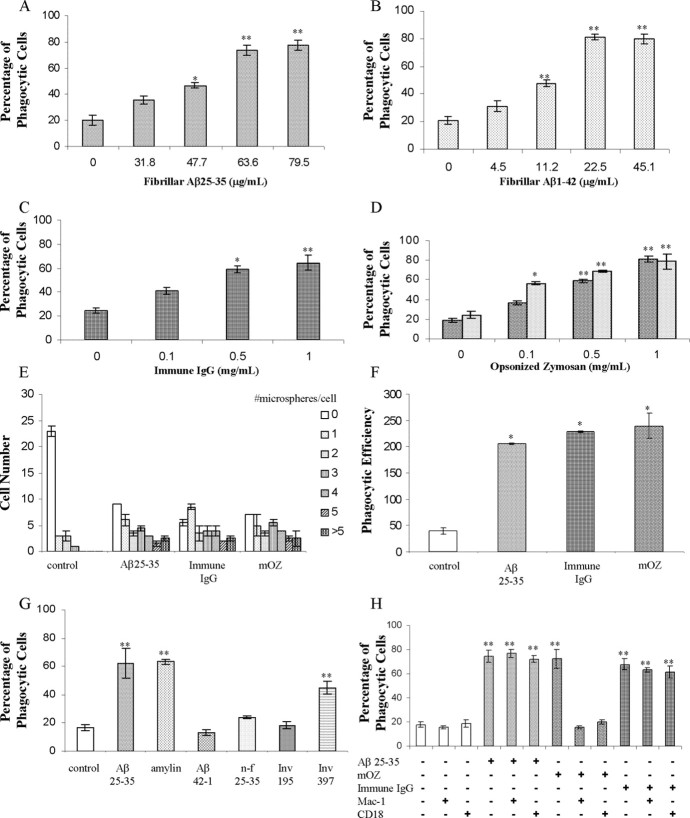
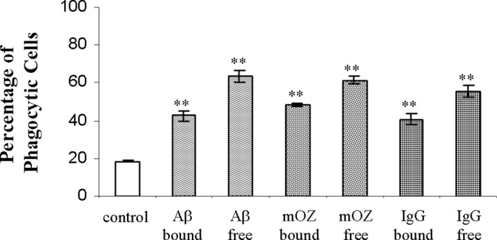
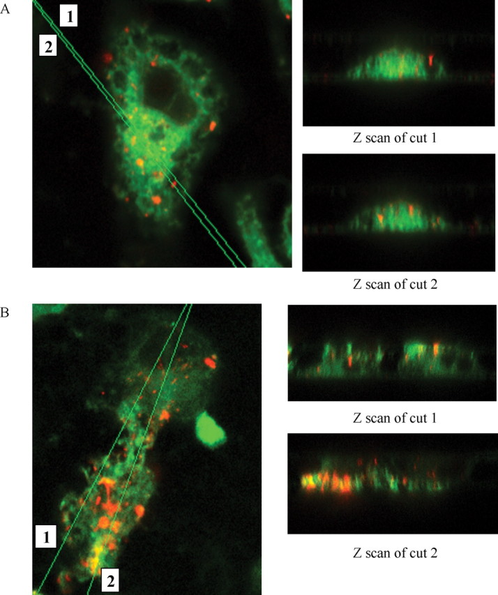
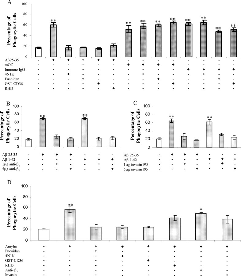
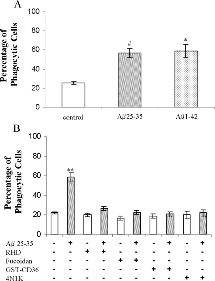
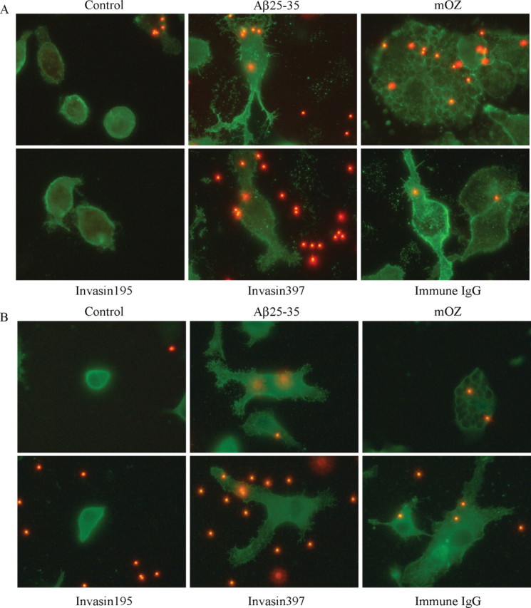
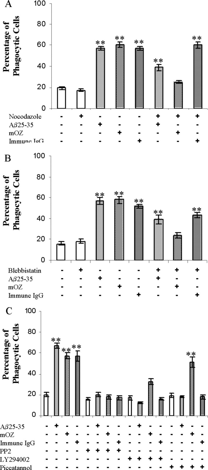
Microglia are the principle immune effector and phagocytic cells in the CNS. These cells are associated with fibrillar beta-amyloid (fAbeta)-containing plaques found in the brains of Alzheimer's disease (AD) patients. The plaque-associated microglia undergo a phenotypic conversion into an activated phenotype and are responsible for the development of a focal inflammatory response that exacerbates and accelerates the disease process. Paradoxically, despite the presence of abundant activated microglia in the brain of AD patients, these cells fail to mount a phagocytic response to Abeta deposits but can efficiently phagocytose Abeta fibrils and plaques in vitro. We report that exposure of microglia to fAbeta in vitro induces phagocytosis through mechanisms distinct from those used by the classical phagocytic receptors, the Ig receptors (FcRgammaI and FcgammaRIII) or complement receptors. Microglia interact with fAbeta through a recently characterized Abeta cell surface receptor complex comprising the B-class scavenger receptor CD36, alpha6beta1 integrin, and CD47 (integrin-associated protein). Antagonists specific for each component of the receptor complex blocks fAbeta-stimulated phagocytosis. These data demonstrated that engagement of this ensemble of receptors is required for induction of phagocytosis. The phagocytic response stimulated by this receptor complex is driven principally by a beta(1) integrin-linked process that is morphologically and mechanistically distinct from the classical type I and type II phagocytic mechanisms. These data provide evidence for phagocytic uptake of fAbeta through a receptor-mediated, nonclassical phagocytic mechanism.


Figures

Figure 1.
Figure 1.
BV-2 cells exhibit ligand-stimulated phagocytosis through classical and nonclassical mechanisms. Dose dependence of phagocytosis stimulated by fAβ25-35 (A), fAβ1-42 (B), immune IgG (C), mouse complement-opsonized zymosan (D) (dark speckles), and rabbit complement-opsonized zymosan (light speckles) in BV-2 cells is shown. BV-2 cells were incubated with the indicated ligand for 30 min before being exposed to microspheres for an additional 30 min. E, The number of microspheres taken up per BV-2 cell in untreated cells or treated with fAβ25-35 (60 μm; 63.6 μg/ml), immune IgG (1 mg/ml), and mouse complement-opsonized zymosan (mOZ) (1 mg/ml) are shown. BV-2 cells were incubated with the indicated ligand for 30 min before the addition of microspheres for 30 min. F, Phagocytic efficiency of cells stimulated with fAβ25-35 (60 μm; 63.6 μg/ml), immune IgG (1 mg/ml), and mouse complement-opsonized zymosan (mOZ) (1 mg/ml). G, BV-2 cells were treated with amylin (10 μm; 39 μg/ml), fAβ25-35 (60 μm; 63.6 μg/ml), invasin397 (Inv 397) (5 μg/ml), Aβ42-1 (5 μm; 22.6 μg/ml), the reverse β amyloid peptide, nonfibrillar (n-f) Aβ25-35 (60 μm; 63.6 μg/ml), and invasin 195 (Inv 195) (5 μg/ml) for 30 min before a 30 min incubation with microspheres. H, Antibodies directed against CR3 [Mac-1 (5 μg/ml) and CD18 (5 μg/ml)] were added to BV-2 cells before exposure to the stimulating ligand. These experiments are representative of three independent studies. *p < 0.01; **p < 0.001 compared with control.
Figure 2.
Figure 2.
Free and immobilized ligands both stimulate phagocytosis. BV-2 cells were exposed to fAβ25-35, immune IgG, or mouse complement-opsonized zymosan (mOZ) either free in the media or immobilized on the surface of the microspheres for 40 min before being scored for ingestion of microspheres. The data shown are representative of three independent experiments. **p < 0.001 compared with control.
Figure 3.
Figure 3.
BV-2 cells phagocytose fibrillar Aβ1-42 labeled with Cy3. Confocal images (100×) of BV-2 cells ingesting Cy3-labeled fAβ1-42 are shown in the x-y plane, as well as Z scans of cuts through the cells. A, BV-2 cells were incubated with Cy3Aβ (40μg/ml) for 30 min before being fixed and stained with DiO (green) to label internal and external cell membranes. B, BV-2 cells were pretreated with unlabeled fAβ25-35 (60 μm) for 30 min before adding Cy3Aβ (40 μg/ml). The ingested Cy3Aβ (red) is trafficked to intracellular vesicles. This experiment is representative of three independent experiments.
Figure 4.
Figure 4.
fAβ elicits phagocytosis through stimulation of the microglial Aβ cell surface receptor complex. A, Antagonists of the microglial Aβ cell surface receptor complex were tested for their ability to inhibit fAβ-mediated phagocytosis. Antagonists of CD47 (100 μm 4N1K), the β1 integrin (100 μg/ml RHD), and scavenger receptors (300 μg/ml fucoidan and 100 nm GST-CD36) were added to BV-2 cells 30 min before stimulation with the indicated ligand. mOZ, Mouse complement-opsonized zymosan. B, C, The anti-β1 antibody as well as invasin 195, a specificβ1 integrin ligand, were added to BV-2 cells 30 min before stimulation with fAβ. D, Antagonists were added to BV-2 for 30 min before amylin stimulation using the same concentrations as in A. These experiments are representative of three independent experiments. *p < 0.01; **p < 0.001 compared with control.
Figure 5.
Figure 5.
fAβ stimulates phagocytosis through the microglial Aβ cell surface receptor complex in primary murine microglia. A, Neonatal primary microglial cells were treated with fAβ25-35 (60 μm; 63.6 μg/ml) and fAβ1-42 (5 μm; 22.6 μg/ml) for 30 min before the addition of microspheres. B, Antagonists to the microglial Aβ cell surface receptor complex were tested in primary microglial cells as described in Figure 4. The data are representative of three independent experiments. *p < 0.01; **p < 0.001; #p < 0.05 compared with control.
Figure 6.
Figure 6.
Cytoskeletal morphology differentiates types of phagocytosis. Phalloidin (A), which stains f-actin, andα-actinin (B) was used as cytoskeletal markers to compare cellular morphologies. BV-2 cells were treated with invasin 195 (1 μg/ml), Aβ25-35 (60 μm; 63.6 μg/ml), invasin397 (1 μg/ml), immune IgG (1 mg/ml), and mouse complement-opsonized zymosan (mOZ) (1 mg/ml) for 30 min before an additional 30 min incubation with microspheres. Images of cells were taken at 100×. These experiments are representative of three independent studies.
Figure 7.
Figure 7.
Cytoskeletal perturbation and signaling inhibitors differentiate types of phagocytosis. A, Nocodazole (5 μg/ml) was added to BV-2 cells to inhibit microtubule dynamics 45 min before stimulation with fAβ25-35 (60 μm; 63.6 μg/ml), immune IgG (1 mg/ml), and mouse complement-opsonized zymosan (mOZ) (1 mg/ml). B, Blebbistatin (100 μm) was added to BV-2 cells 30 min before stimulation with the indicated ligand. C, Piceatannol (10 μm), a Syk inhibitor, PP2 (10 μm), a Src inhibitor, and LY294002 (10 μm), a PI3K inhibitor, were added to BV-2 cells 30 min before stimulation with the indicated ligand. The data are representative of five independent experiments. **p < 0.001 compared with control.

References

    1. Aderem A, Underhill DM (1999) Mechanisms of phagocytosis in macrophages. Annu Rev Immunol 17: 593-623. - PubMed
    1. Allen LA, Aderem A (1996) Molecular definition of distinct cytoskeletal structures involved in complement- and Fc receptor-mediated phagocytosis in macrophages. J Exp Med 184: 627-637. - PMC - PubMed
    1. Ard MD, Cole GM, Wei J, Mehrle AP, Fratkin JD (1996) Scavenging of Alzheimer's amyloid beta-protein by microglia in culture. J Neurosci Res 43: 190-202. - PubMed
    1. Bamberger ME, Harris ME, McDonald DR, Husemann J, Landreth GE (2003) A cell surface receptor complex for fibrillar beta-amyloid mediates microglial activation. J Neurosci 23: 2665-2674. - PMC - PubMed
    1. Bard F, Cannon C, Barbour R, Burke RL, Games D, Grajeda H, Guido T, Hu K, Huang J, Johnson-Wood K, Khan K, Kholodenko D, Lee M, Lieberburg I, Motter R, Nguyen M, Soriano F, Vasquez N, Weiss K, Welch B, Seubert P, Schenk D, Yednock T (2000) Peripherally administered antibodies against amyloid beta-peptide enter the central nervous system and reduce pathology in a mouse model of Alzheimer disease. Nat Med 6: 916-919. - PubMed

Publication types