. 2004 Nov 3;24(44):9903-13.
doi: 10.1523/JNEUROSCI.1713-04.2004.

Late calcium EDTA rescues hippocampal CA1 neurons from global ischemia-induced death

Affiliations

Late calcium EDTA rescues hippocampal CA1 neurons from global ischemia-induced death

Agata Calderone et al. J Neurosci. .

Abstract

Transient global ischemia induces a delayed rise in intracellular Zn2+, which may be mediated via glutamate receptor 2 (GluR2)-lacking AMPA receptors (AMPARs), and selective, delayed death of hippocampal CA1 neurons. The molecular mechanisms underlying Zn2+ toxicity in vivo are not well delineated. Here we show the striking finding that intraventricular injection of the high-affinity Zn2+ chelator calcium EDTA (CaEDTA) at 30 min before ischemia (early CaEDTA) or at 48-60 hr (late CaEDTA), but not 3-6 hr, after ischemia, afforded robust protection of CA1 neurons in approximately 50% (late CaEDTA) to 75% (early CaEDTA) of animals. We also show that Zn2+ acts via temporally distinct mechanisms to promote neuronal death. Early CaEDTA attenuated ischemia-induced GluR2 mRNA and protein downregulation (and, by inference, formation of Zn2+-permeable AMPARs), the delayed rise in Zn2+, and neuronal death. These findings suggest that Zn2+ acts at step(s) upstream from GluR2 gene downregulation and implicate Zn2+ in transcriptional regulation and/or GluR2 mRNA stability. Early CaEDTA also blocked mitochondrial release of cytochrome c and Smac/DIABLO (second mitochondria-derived activator of caspases/direct inhibitor of apoptosis protein-binding protein with low pI), caspase-3 activity (but not procaspase-3 cleavage), p75NTR induction, and DNA fragmentation. These findings indicate that CaEDTA preserves the functional integrity of the mitochondrial outer membrane and arrests the caspase death cascade. Late injection of CaEDTA at a time when GluR2 is downregulated and caspase is activated inhibited the delayed rise in Zn2+, p75NTR induction, DNA fragmentation, and cell death. The finding of neuroprotection by late CaEDTA administration has striking implications for intervention in the delayed neuronal death associated with global ischemia.


Figures
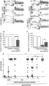
Figure 2.
Figure 2.
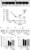
Early and late CaEDTA afford protection against ischemia-induced neuronal death. Toluidine blue-stained coronal brain sections at the level of the dorsal hippocampus from control (n = 3; a, b) and experimental animals subjected to CaEDTA injection (c, d), global ischemia (e, f), or to CaEDTA (300 mm, i.c.v.), followed by ischemia (g, h) and killed 5 d after surgery. Brain sections from sham (n = 3; i, j) and experimental animals subjected to CaEDTA injection (k, l), ischemia (n = 4; m, n), or to ischemia, followed by CaEDTA at 60 hr after ischemia (n = 6; o-r) and killed 7 d after ischemia. Late CaEDTA afforded robust neuroprotection is some (q, r), but not other (o, p), animals. s, t, Quantitation of cell counts from sections such as those shown in a-h (s) and i-r (t). u, Summary data for CaEDTA injections given at times ranging from 30 min before ischemia to 72 hr after ischemia. Global ischemia was induced in gerbils by BCCO (5 min). Animals were killed at 7 d after ischemia, and brain sections at the level of the dorsal hippocampus were scored for neuronal damage according to Pulsinelli and Brierley (1979). CaEDTA afforded neuroprotection in a substantial number of animals when administered 30 min before ischemia or at 48 or 60 hr after ischemia but was ineffective when administered at 3 or 6 hr after ischemia. Statistical significance of changes in neuronal counts (s, t) was assessed by means of Student's unpaired t test and of changes in neuronal damage scores (u) by means of ANOVA (*p < 0.05; **p < 0.01; ***p < 0.001). Error bars represent SEM. Scale bars: (in a, i) a, c, e, g, i, k, m, o, q, 500 μm; (in b, j) b, d, f, h, j, l, n, p, r, 50 μm. so, Stratum oriens; sp, stratum pyramidale; sr, stratum radiatum.
Figure 1.
Figure 1.
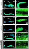
Global ischemia elicits a highly delayed rise in Zn2+ in selectively vulnerable CA1 neurons. Zn2+ fluorescence in TSQ-stained coronal brain sections from sham (a-d) and experimental animals subjected to global ischemia (e-l) or to CaEDTA, followed by global ischemia (k, l). In control hippocampus, TSQ labeling revealed intense fluorescence in the mossy fiber axon terminals of dentate granule neurons in the hilus (h) and stratum lucidum of CA3 (slu) and faint fluorescence in the stratum radiatum and stratum oriens of CA1 (a, b). CaEDTA injection at 30 min before surgery did not detectably alter the pattern of Zn2+ fluorescence in sham-operated control animals, assessed at 72 hr after surgery (c, d). Global ischemia induced a pronounced increase in Zn2+ fluorescence in the cell bodies of scattered hilar neurons, evident at 24 hr after insult (e, f). At 48 hr, Zn2+ fluorescence was visible in CA3a pyramidal neurons, extending into the CA1/CA3 transition zone, but was not visible in CA1 (g, h). At 72 hr, Zn2+ fluorescence was pronounced in cell bodies of CA1 pyramidal neurons [i (arrows), j]. Injection of CaEDTA 30 min before ischemia did not affect the increase in Zn2+ in the transition zone but attenuated the late rise in Zn2+ fluorescence in the CA1 (k, l). Scale bars: (in a) a, c, e, g, i, k, 400 μm; (in b) b, d, f, h, j, l, 80 μm. so, Stratum oriens; sp, stratum pyramidale; sr, stratum radiatum.
Figure 3.
Figure 3.
Early CaEDTA attenuates ischemia-induced suppression of GluR2 mRNA and protein in CA1. a, b, Representative film autoradiograms of GluR2 mRNA expression detected by in situ hybridization in the hippocampus of control and experimental animals subjected to global ischemia at 48 hr (n = 3), 72 hr (n = 4), and 5 d (n = 3) after reperfusion (a) or to pretreatment with CaEDTA (300 mm, i.c.v.), followed by ischemia at 48 hr (n = 4), 72 hr (n = 4), and 5 d after reperfusion (n = 4) (b). c, Quantitative analysis of data such as those in a, b. Mean optical densities were normalized to the corresponding control value for a given region. Ischemia caused a loss of GluR2 mRNA in CA1 (but not DG or CA3), evident at 48 hr. CaEDTA pretreatment did not alter ischemia-induced downregulation of GluR2 mRNA (48 hr) but accelerated its recovery to control values (72 hr and 5 d). d, e, Representative Western blots probed with a monoclonal antibody against a sequence within the N-terminal domain of the GluR2 subunit. f, g, Relative GluR2 subunit abundance for protein samples isolated from the CA1 (f) and DG (g) of control (n = 4) and experimental animals at 48 hr after CaEDTA injection (n = 4), 48 hr (n = 4) and 72 hr (n = 3) after ischemia, and at 48 hr (n = 4) and 72 hr (n = 4) after CaEDTA injection, followed by ischemia. GluR2 abundance was determined from band densities for GluR2 after normalization to the band densities for actin, which served as a loading control. Relative GluR2 subunit abundance was markedly decreased in CA1 (but not DG) at 48 and 72 hr after ischemia. CaEDTA pretreatment attenuated GluR2 downregulation, assessed at 72 hr and 5 d. Bars are means ± SEM. Statistical significance was assessed by means of Student's unpaired t test (*p < 0.05; **p < 0.01; ***p < 0.001).
Figure 4.
Figure 4.
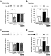
Early CaEDTA blocks the ischemia-induced mitochondrial release of cytochrome c (Cyt c) and Smac/DIABLO. Representative Western blots and relative abundance of cytochrome c (a, b) and Smac/DIABLO (c, d) in the mitochondrial (a, c) and cytosolic (b, d) fractions of protein samples isolated from control and experimental animals subjected to CaEDTA injection, ischemia, or CaEDTA (300 mm, i.c.v.; 30 min before ischemia), followed by ischemia and killed at 12 hr after reperfusion. Westerns were probed with anti-cytochrome c antibody (a, b) or anti-Smac/DIABLO antibody (c, d). A single injection of CaEDTA significantly reduced the mitochondrial and cytosolic levels of cytochrome c(a, b) and cytosolic levels of Smac/DIABLO (d) in control animals and greatly attenuated the ischemia-induced increase in cytosolic cytochrome c (b) and Smac/DIABLO (d). Mean band densities for cytochrome c or Smac/DIABLO in protein samples from the CA1 of experimental animals were normalized to the corresponding values for samples from control animals. Statistical significance was assessed by ANOVA, followed by Newman-Keuls test (**p < 0.01). Error bars represent SEM.
Figure 5.
Figure 5.
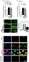
Early CaEDTA acts downstream of caspase-3 cleavage to block caspase-3 activity, p75NTR induction, and TUNEL. Representative Western blots and relative abundance of procaspase-3 (a) and caspase-3 (b) in protein samples isolated from control (Contl) and experimental animals subjected to ischemia or CaEDTA (300 mm, i.c.v.; 30 min before ischemia), followed by ischemia and killed at 24 hr after reperfusion. Westerns were probed with an anti-caspase-3 antibody. Global ischemia induced an upregulation of procaspase-3 (a) and promoted its proteolytic processing to generate cleaved (activated) caspase-3 (b). CaEDTA did not significantly alter the ischemia-induced upregulation of procaspase-3 (a) or the appearance of caspase-3 (b). Mean band densities for procaspase-3 and cleaved (activated) caspase-3 in protein samples from the CA1 of experimental animals were normalized to the corresponding values for samples from control animals. Representative brain sections at the level of the dorsal hippocampus from animals subjected to sham operation (control; c, d), global ischemia (e, f), or CaEDTA (300 mm, i.c.v.; 30 min before ischemia), followed by ischemia and killed at 24 hr (g, h), labeled with FAM-DEVD-FMK, a fluorescein-tagged analog of zDEVD-FMK, a potent inhibitor of activated caspases. c, d, Caspase-3 activity was low in control brain. e, f, Global ischemia induced caspase-3 activity in the CA1 pyramidal cell layer, evident at 24 hr (g, h). i, Summary of data for animals subjected to sham operation, ischemia, or CaEDTA, followed by ischemia. Scale bars, 400 μm (low magnification); 50 μm (high magnification). Error bars represent SEM.j-u, Triple labeling of representative brain sections with the nuclear stain Hoechst 33342 (j, m, p, s), an anti-p75NTR antibody (k, n, q, t), and TUNEL (l, o, r, u) in the CA1 pyramidal layer of control (j, l) and experimental gerbils subjected to global ischemia (m-r) or to CaEDTA (300 mm, i.c.v.; 30 min before ischemia), followed by ischemia and killed at 72 hr (s-u). p is the super imposition of m and n.q is a superimposition of m and o. r is a superimposition of n and o. Indicated boxes are enlarged in p-r. p75NTR immunolabeling visualized in red (Texas Red), TUNEL in green (fluorescein), and Hoechst in blue. so, Stratum oriens; sp, stratum pyramidale; sr, stratum radiatum. Global ischemia induced p75NTR expression and TUNEL positivity in CA1 pyramidal neurons; CaEDTA pretreatment reduced p75NTR immunolabeling and TUNEL. Scale bars: (in r) p-r, 10 μm; (in u) j-o and s-u, 60 μm. Statistical significance in a, b, i was assessed by ANOVA, followed by Newman-Keuls test (*p < 0.05; **p < 0.01).
Figure 6.
Figure 6.
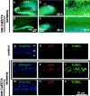
Late CaEDTA blocks the ischemia-induced rise in Zn2+ and late-stage apoptosis in CA1 neurons. Zn2+ fluorescence in TSQ-stained coronal brain sections from experimental animals subjected to global ischemia (a-c) or to global ischemia, followed by CaEDTA (300 mm, i.c.v. at 60 hr) and killed at 72 hr after reperfusion (d-f). Global ischemia induced a pronounced increase in Zn2+ fluorescence in the cell bodies of scattered hilar neurons (a) and CA3 pyramidal neurons (b). At 72 hr, Zn2+ fluorescence was pronounced in cell bodies of CA1 pyramidal neurons (c). Late injection of CaEDTA prevented the rise in Zn2+ fluorescence in CA1 neurons. g-o, Representative brain sections at the level of the dorsal hippocampus labeled with the nuclear stain Hoechst 33342 (g, j, m), an anti-p75NTR antibody (h, k, n), and TUNEL (i, l, o) from control (g-i) and experimental gerbils subjected to global ischemia (j-l) or to global ischemia followed by CaEDTA (300 mm, i.c.v. at 60 hr) and killed at 72 hr after reperfusion (m-o). p75NTR immunolabeling visualized in red (Texas Red), TUNEL reaction in green (fluorescein), and Hoechst-stained nuclei in blue. so, Stratum oriens; sp, stratum pyramidale; sr, stratum radiatum. Coincidence of p75NTR immunolabeling and TUNEL positivity was observed in neurons throughout the CA1 pyramidal cell layer; CaEDTA injection at 60 hr after reperfusion markedly reduced p75NTR expression and TUNEL. Scale bars: a, d, 400 μm; b, c, e, f, 80 μm; g-o, 60 μm.

Similar articles

Cited by

References

    1. Aizenman E, Stout AK, Hartnett KA, Dineley KE, McLaughlin B, Reynolds IJ (2000) Induction of neuronal apoptosis by thiol oxidation: putative role of intracellular zinc release. J Neurochem 75: 1878-1888. - PubMed
    1. Bers DM, Patton CW, Nuccitelli R (1994) A practical guide to the preparation of Ca2+ buffers. Methods Cell Biol 40: 3-29. - PubMed
    1. Brene S, Messer C, Okado H, Hartley M, Heinemann SF, Nestler EJ (2000) Regulation of GluR2 promoter activity by neurotrophic factors via a neuron-restrictive silencer element. Eur J Neurosci 12: 1525-1533. - PubMed
    1. Bresink I, Ebert B, Parsons CG, Mutschler E (1996) Zinc changes AMPA receptor properties: results of binding studies and patch clamp recordings. Neuropharmacology 35: 503-509. - PubMed
    1. Calderone A, Jover T, Noh K-M, Tanaka H, Yokota H, Lin Y, Grooms S, Regis R, Bennett MVL, Zukin RS (2003) Ischemic insults derepress the gene silencer REST in neurons destined to die. J Neurosci 23: 2112-2121. - PMC - PubMed

Publication types

MeSH terms