. 2004 Nov 3;24(44):10003-12.
doi: 10.1523/JNEUROSCI.2114-04.2004.

p53 activation domain 1 is essential for PUMA upregulation and p53-mediated neuronal cell death

Affiliations

p53 activation domain 1 is essential for PUMA upregulation and p53-mediated neuronal cell death

Sean P Cregan et al. J Neurosci. .

Abstract

The p53 tumor suppressor gene has been implicated in the regulation of apoptosis in a number of different neuronal death paradigms. Because of the importance of p53 in neuronal injury, we questioned the mechanism underlying p53-mediated apoptosis in neurons. Using adenoviral-mediated gene delivery, reconstitution experiments, and mice carrying a knock-in mutation in the endogenous p53 gene, we show that the transactivation function of p53 is essential to induce neuronal cell death. Although p53 possesses two transactivation domains that can activate p53 targets independently, we demonstrate that the first activation domain (ADI) is required to drive apoptosis after neuronal injury. Furthermore, the BH3-only proteins Noxa and PUMA exhibit differential regulation by the two transactivation domains. Here, we show that Noxa can be induced by either activation domain, whereas PUMA induction requires both activation domains to be intact. Unlike Noxa, the upregulation of PUMA alone is sufficient to induce neuronal cell death. We demonstrate, therefore, that the first transactivation domain of p53 is indispensable for the induction of neuronal cell death.


Figures

Figure 1.
Figure 1.
Schematic representation of p53 constructs. Various p53 constructs were used to determine the importance of each functional domain for neuronal apoptosis. The p53ΔI construct contains a deletion of the conserved box 1 sequence, which abolishes MDM2 binding without affecting DNA-binding activity. The p53Δ22/23 construct contains two inactivating point mutations within the first transactivation domain (ADI); residues 22 and 23 were mutated from Leu and Trp to Glu and Ser, respectively. p53Δ53/54 has mutations at residues 53 and 54 from Trp and Glu to Phe and Ser, respectively, which inactivates the second transactivation domain (ADII). The p53DM contains both transactivation domain mutations (p53-Δ22/23 and p53-Δ53/54). p53ΔPro is a deletion of the proline-rich region of the p53 protein, the P53-Δ173L mutant has a point mutation at residue 173 to Leu, and p53ΔV is a deletion of the conserved box V sequence of p53, resulting in inactivation of DNA binding.
Figure 2.
Figure 2.
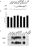
P53 mutant proteins are stable, expressed at comparable levels, and retain the appropriate protein conformation. A, Western blot showing comparable expression levels of p53 mutant proteins. CGNs were infected at 25 MOI with the indicated Ad-p53 construct, and protein lysates were collected at 36 hr after infection (30 μg protein/lane). B, CGNs were infected with the indicated Ad-p53 construct at 25 MOI. After 36 hr, neurons were fixed and immunostained for p53 and counterstained with Hoechst. Data represent the mean and SD of three independent experiments. C, EMSA. Protein was extracted from CGNs 36 hr after infection with the indicated Ad-p53 constructs. p53-binding activity to the APAF1 and p21 p53 response elements were assayed by EMSA. Binding reactions were performed with neuronal extracts (10-20 μg protein) and the indicated oligonucleotides in the presence of p53 antibody (Ab1). To control for binding specificity, a 100-fold excess of unlabeled oligonucleotide was added to the binding reaction and incubated for 20 min before the addition of labeled probe. All p53 constructs tested efficiently bound DNA, with the exception of ΔV, which inactivates DNA binding (n = 3).
Figure 3.
Figure 3.
p53 mutant proteins localize to the nucleus. Cortical neurons were infected with Ad-p53WT or Ad-p53DM at 25 MOI. After 48 hr, neurons were fixed and immunostained for p53 (A, B) and/or mitochondrial-specific CoxIV (B) or counterstained with Hoechst (A). p53 remained localized to the nucleus even under conditions of cell death, as denoted by condensed nuclei (b).
Figure 4.
Figure 4.
Transactivation domain 1 of p53 is essential for the induction of neuronal cell death. CGNs were infected with wild-type Ad-p53 or p53 mutant constructs at 25 MOI. A, LIVE/DEAD viability/cytotoxicity assay (Molecular Probes) was preformed at 24, 48, 72, and 96 hr after infection. B, Photomicrographs of LIVE/DEAD cell assay. Scale bar, 100 μm. C, Caspase-3 activity was measured at 48 and 72 hr by DEVD-AFC cleavage. D, The fraction of TUNEL-positive cells was measured 72 hr after infection. Data represent the mean and SD of five independent experiments (n = 5).
Figure 5.
Figure 5.
Reconstitution of responsiveness to DNA damage-induced apoptosis in p53-deficient neurons. Cortical neurons obtained from p53-deficient mice or wild-type littermates were infected with Ad-p53 or Ad-p53 mutant constructs and 16 hr later were challenged with 10 μm camptothecin. The reconstitution of apoptotic cell death was determined by LIVE/DEAD assay 24 hr after treatment. *p < 0.05 by two-way ANOVA compared with wild type, followed by t test. Data represent the mean and SD of three independent experiments (n = 3). Ctrl, Control.
Figure 6.
Figure 6.
p53 mutants cause differential upregulation of target genes in CGNs. Total RNA was extracted from CGNs 40 hr after infection with Ad-p53 or Ad-p53 mutant constructs as indicated and analyzed for Noxa, Apaf-1, PUMA, and Perp or S12 expression using semiquantitative RT-PCR. CTRL, Control.
Figure 7.
Figure 7.
Transactivation domain 1 of p53 is essential for the induction of neuronal cell death in vivo. A, Cortical neurons obtained from QS mice, p53-deficient mice, or wild-type littermates were treated with 10 μm camptothecin, and cell survival was determined by LIVE/DEAD assay at the indicated times. Cell death is reported as a percentage of corresponding untreated control cultures. Data represent the mean and SD from three independent experiments (n = 3). B, Total RNA from cortical neurons from CD1 mice treated with camptothecin was collected and analyzed at the indicated times for Noxa, PUMA, and S12 expression using semiquantitative RT-PCR. C, Cortical neurons from QS mice, p53-deficient mice, or wild-type littermates were treated with 10 μm camptothecin, and after 9 hr, total RNA was collected and analyzed for Noxa, PUMA, and S12 expression using semiquantitative RT-PCR.
Figure 8.
Figure 8.
Upregulation of Noxa and PUMA mRNA in p53-mediated neuronal cell death. Western blots showing efficient transduction of Ad-Noxa3XFlag and AD-PumaHA are shown. A, CGNs were infected with Ad-Noxa3XFlag or Ad-GFP at 50 MOI. Protein lysates were collected 48 hr after infection and were blotted for Noxa expression with an antibody against Flag or for actin as a loading control. B, HEK 293 cells were infected with Ad-PumaHA or Ad-GFP at 25 MOI. Protein lysates were collected 12 hr after infection and were blotted for PUMA expression with an antibody against HA or for actin as a loading control. C, Neurons were infected with Ad-Noxa-Flag or Ad-PumaHA at 50 MOI. After 24 hr, neurons were fixed and immunostained for Flag or HA and counterstained with Hoechst. Scale bar, 25 μm. Figures are a representative of three independent experiments (n = 3).
Figure 9.
Figure 9.
Forced expression of PUMA, but not Noxa, is sufficient to induce neuronal apoptosis. CGNs were infected with Ad-Noxa3XFlag (A), Ad-PumaHA (B), or Ad-GFP control at the indicated MOI. Neuronal survival was determined at the indicated times by MTT assay. Survival is measured as a percentage of Ad-GFP-treated control cells. C, CGNs were infected with Ad-Noxa3XFlag or Ad-PumaHA at 50 MOI. Neuronal survival was determined at the indicated times by MTT assay. Survival was measured as a percentage of Ad-GFP-treated control cells. Data represent the mean and SD from three independent experiments (n = 3).
Figure 10.
Figure 10.
Upregulation of PUMA is essential for the induction of p53-mediated neuronal cell death. CGNs obtained from PUMA-deficient mice or control littermates were infected with wild-type Ad-p53, p53ΔDM, or GFP constructs at 15 MOI. LIVE/DEAD viability/cytotoxicity assay (Molecular Probes) was preformed at 48 hr (A), 72 hr (B), and 96 hr (C) after infection. Data represent the mean and SD of three independent experiments (n = 3).

References

    1. Attardi LD, Lowe SW, Brugarolas J, Jacks T (1996) Transcriptional activation by p53, but not induction of the p21 gene, is essential for oncogene-mediated apoptosis. EMBO J 15: 3693-3701. - PMC - PubMed
    1. Attardi LD, Reczek EE, Cosmas C, Demicco EG, McCurrach ME, Lowe SW, Jacks T (2000) PERP, an apoptosis-associated target of p53, is a novel member of the PMP-22/gas3 family. Genes Dev 14: 704-718. - PMC - PubMed
    1. Banasiak KJ, Haddad GG (1998) Hypoxia-induced apoptosis: effect of hypoxic severity and role of p53 in neuronal cell death. Brain Res 797: 295-304. - PubMed
    1. Baptiste N, Friedlander P, Chen X, Prives C (2002) The proline-rich domain of p53 is required for cooperation with anti-neoplastic agents to promote apoptosis of tumor cells. Oncogene 21: 9-21. - PubMed
    1. Bennett M, Macdonald K, Chan SW, Luzio JP, Simari R, Weissberg P (1998) Cell surface trafficking of Fas: a rapid mechanism of p53-mediated apoptosis. Science 282: 290-293. - PubMed

Publication types

Substances