. 2004 Nov 24;23(23):4583-94.
doi: 10.1038/sj.emboj.7600475. Epub 2004 Nov 4.

Axin stimulates p53 functions by activation of HIPK2 kinase through multimeric complex formation

Affiliations

Axin stimulates p53 functions by activation of HIPK2 kinase through multimeric complex formation

Yanning Rui et al. EMBO J. .

Abstract

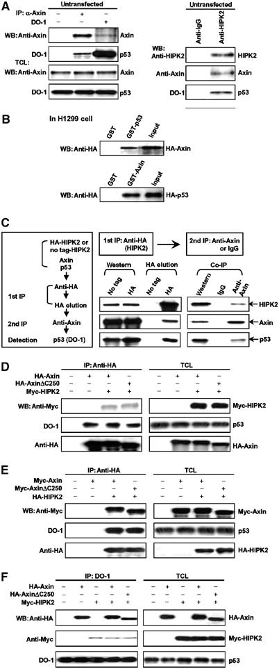
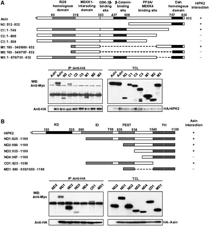
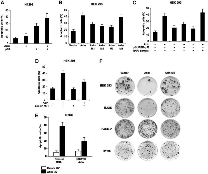
Axin and p53 are tumor suppressors, controlling cell growth, apoptosis, and development. We show that Axin interacts with homeodomain-interacting protein kinase-2 (HIPK2), which is linked to UV-induced p53-dependent apoptosis by interacting with, and phosphorylating Ser 46 of, p53. In addition to association with p53 via HIPK2, Axin contains a separate domain that directly interacts with p53 at their physiological concentrations. Axin stimulates p53-dependent reporter transcription in 293 cells, but not in 293T, H1299, or SaOS-2 cells that are defective in p53 signaling. Axin, but not AxindeltaHIPK2, activates HIPK2-mediated p53 phosphorylation at Ser 46, facilitating p53-dependent transcriptional activity and apoptosis. Specific knockdown of Axin by siRNA reduced UV-induced Ser-46 phosphorylation and apoptosis. Kinase-dead HIPK2 reduced Axin-induced p53-dependent transcriptional activity, indicating that Axin stimulates p53 function through HIPK2 kinase activity. Interestingly, HIPK2deltaAxin that lacks its Axin-binding region acts as a dominant-positive form in p53 activation, suggesting that the Axin-binding region of HIPK2 is a putative autoinhibitory domain. These results show that Axin acts as a tumor suppressor by facilitating p53 function through integration of multiple factors.


Figures

Figure 1
Figure 1
Interaction of Axin with HIPK2 and p53 in vivo and in vitro. (A) Axin and p53 in untransfected 293T cells were separately immunoprecipitated with rabbit anti-Axin that was raised against aa 348–500 of mouse Axin and anti-p53 antibody DO-1, respectively (left panel). The control IgG used was from rabbit. Detection of Axin and p53 in the immunoprecipitates and total cell lysates (TCL) was carried out using anti-Axin and DO-1 antibodies. For analysis of HIPK2 interaction with Axin and p53, we carried out immunoprecipitation with goat anti-HIPK2 antibody or control goat IgG and detected copresence of Axin and p53 in its immunoprecipitate (right panel). (B) GST pulldown assay to evaluate Axin interaction with p53 in vitro. The ability of GST-p53 to retain Axin present in the cell lysate from H1299 cells transfected with pCMV-HA-Axin was analyzed by Western blotting with anti-HA following SDS–PAGE (top panel). Bottom: The ability of GST-Axin to pull down HA-p53 in the cell lysate. In either case, GST alone did not interact with p53 or Axin. Input represents 1/4 of that used for GST pulldown. (C) Two-step co-immunoprecipitation of the complex containing Axin, HIPK2, and p53. The procedures of the two-step co-immunoprecipitation are outlined in the box on the left, according to Harada et al (2003). HEK 293T cells were transfected with plasmids expressing Myc-Axin and HA-HIPK2 (or untagged HIPK2 as control). The first immunoprecipitation was performed using anti-HA and Protein A/G-agarose beads. The complex was eluted with 3 × HA-tag, followed by the second step of co-immunoprecipitation with anti-Axin or control IgG. Protein samples from each step were analyzed by Western blotting separately with anti-Axin, anti-HIPK2, and anti-p53 (DO-1). (D–F) HIPK2 and p53 form a complex on the monomeric Axin. AxinΔC250, which lacks the DIX domain and cannot form homodimer, was used in this experiment. (D) HA-Axin and Myc-HIPK2 were transfected into HEK 293T cells and immunoprecipitation was performed with anti-HA antibody. HIPK2 and endogenous p53 were detected in the Axin immunoprecipitate by Western blotting with anti-Myc and DO-1 (anti-p53) antibody, respectively. Protein levels of Axin, HIPK2, and p53 in the total cell lysates (TCL) were determined by anti-HA, anti-Myc, and DO-1, respectively (on the right). (E) Axin and p53 are copresent in HIPK2 immunoprecipitates. HA-HIPK2 and Myc-Axin were cotransfected into HEK 293T cells and HIPK2 was immunoprecipitated with anti-HA. Co-precipitated endogenous p53 and Myc-Axin were detected with DO-1 and anti-Myc antibody, respectively. (F) Endogenous p53 forms a complex with Axin and HIPK2. Immunoprecipitation was carried out with DO-1. Axin and HIPK2 were detected with anti-HA and anti-Myc, respectively.
Figure 2
Figure 2
Determination of domains for mutual interaction between Axin and HIPK2. (A) Determination of HIPK2-binding sites on Axin. Schematic diagrams (on the top) depict different Axin deletion constructs used in the domain mapping experiments. The different Axin deletion constructs were cotransfected with the blank vector (first lane) or the full-length HA-HIPK2 into HEK 293T cells. Cells were then subjected to lysis and immunoprecipitation, followed by Western blotting with different antibodies indicated. (B) Identification of HIPK2 sequence critical for Axin binding. On the top are schematic diagrams depicting different HIPK2 deletion mutants used in the domain mapping experiments. HEK 293T cells were transfected with HA-Axin and Myc-tagged HIKP2 deletion constructs. Cell lysates were immunoprecipitated with anti-HA antibody. The immunoprecipitates and cell lysates were then analyzed by Western blotting separately using anti-HA for HA-Axin, and anti-Myc for Myc-HIPK2 deletion constructs.
Figure 3
Figure 3
Identification of critical regions of Axin and p53 for their complex formation. (A) Endogenous p53 directly interacts with Axin via a domain distinct from the HIPK2-binding site. Schematic diagrams depict different Axin deletion mutants used in the domain mapping experiments. Different HA-tagged Axin deletion constructs were transiently transfected into HEK 293T cells. Cell lysates were immunoprecipitated with anti-p53 antibody DO-1, followed by immunoblotting using anti-HA for Axin deletion mutants and DO-1 for endogenous p53. (B) Determination of Axin-binding sites in p53. Schematic diagrams depict different p53 deletion mutants used in the domain mapping experiments. HEK 293T cells were transfected with HA-Axin and different Myc-tagged p53 deletion constructs as indicated. Cell lysates were immunoprecipitated with anti-HA antibody. The immunoprecipitates and cell lysates were then analyzed by Western blotting separately using anti-HA for HA-Axin and anti-Myc for Myc-p53 proteins.
Figure 4
Figure 4
Axin enhances p53-dependent transcriptional activity. PathDetect p53-Luc reporter (A–E) or Mdm2 reporter (F) was transfected together with different Axin or p53 constructs into HEK 293, H1299, and SaOS-2 as indicated. Western blotting of expressed proteins was performed to indicate similar expression levels (insets). In each transfection, an equal amount of GFP expression plasmid was included as internal control and the GFP protein level in each cell lysate was probed with anti-GFP (Molecular Probes) as loading control (l.c.). All transfections were performed in duplicate and the data are means±s.d. of five independent experiments after normalizing luciferase activity from the vector control to 1. (A) Axin can activate p53 transactivation in a dose-dependent manner. Axin (0, 0.5, 1.0, 2.0, and 3.0 μg) and 0.5 μg of the PathDetect p53-Luc reporter (Stratagene) were cotransfected in HEK 293 cells to test the effect of Axin on p53-dependent transcriptional activity. (B) Axin mutants defective in p53 binding (M4), HIPK2 binding (M3), or binding to both p53 and HIPK2 (M9) did not stimulate p53 transactivation except M8, which contains both the p53- and HIPK2-binding sites. (C) The DNA-binding-defective p53 mutant p53-R175H negatively affects Axin-stimulated p53 transcriptional activity. HEK 293 cells were transfected with the p53-Luc reporter in different combinations of an empty vector, Axin, and p53-R175H, as indicated. (D) Axin depends on p53 for stimulation of reporter transcriptional activity in p53−/− H1299 cells. Flag-Axin failed to activate the p53 reporter in H1299 cells; in the presence of wild-type p53 expressed under the control of pCMV5 vector (1 ng each), Axin further stimulates the reporter activity in a dose-dependent manner. (E) HA-Axin activates p53-dependent transcription in p53−/− SaOS-2 cells in a similar manner to that described in panel D. (F) An Mdm2 luciferase reporter was cotransfected with increasing amounts of Axin into HEK 293 cells.
Figure 5
Figure 5
Axin and HIPK2 synergistically enhance p53-Luc reporter activity. HEK 293 cells were transfected with expression plasmids in different combinations as indicated. Insets show similar expression levels of relevant transfected proteins. Measurement and data presentation are as described in the legend to Figure 4. (A) Axin and HIPK2 mutually enhance stimulation of p53-Luc transcriptional activity. (B, C) Dominant-negative mutant of HIPK2 (HIPK2-K221R (B)) and siRNA (pSUPER-HIPK2) against HIPK2 (C) diminished Axin activation of p53-Luc reporter gene transcription. It is to be noted that the luciferase activity from cells transfected with HIPK2-K221R alone (bar 3 in panel B) was normalized to 1. RNAi control is expressed from pSUPER that contains an insert with multiple variations to the HIPK2 siRNA. (D) The siRNA against Axin, but not control siRNA, significantly diminished the activation of p53 by HIPK2. (E) AxinΔHIPK2 defective in HIPK2 binding drastically reduced p53-Luc activation by HIPK2. Wild-type (WT) Axin alone was transfected as a positive control for AxinΔHIPK2. (F) HIPK2 and HIPK2ΔAxin defective in Axin binding were separately transfected into HEK 293 cells, and p53 luciferase activity was measured. HIPK2ΔAxin behaved as a dominant positive mutant as it stimulated the reporter activity to a greater extent than the wild-type HIPK2.
Figure 6
Figure 6
Axin stimulates HIPK2-mediated p53 phosphorylation at Ser 46. (A) Axin stimulates p53 phosphorylation in a dose-dependent manner. Increasing amounts of HA-Axin were transfected in H1299 cells together with Myc-p53, and as an internal control GFP-expressing vector pEGFPN1. Cell lysates were immunoprecipitated with anti-Myc antibody, followed by Western blotting with anti-p53 (FL393), anti-HA (Axin), anti-phospho-Ser 46-p53, and anti-GFP. (B) H1299 cells were transfected with HA-Axin, Flag-HIPK2, Myc-p53, and GFP-expressing vector pEGFPN1 as indicated. Cell lysates were immunoprecipitated with anti-Myc antibody. The immunoprecipitates and total cell lysates were then analyzed by immunoblotting separately using anti-p53 (FL393) for p53, anti-HA for Axin, anti-Flag for HIPK2, anti-phospho-Ser 46 for the level of Ser-46-phosphorylated p53, and anti-GFP that serves as a control for similar transfection efficiency. (C) HIPK2 siRNA diminished Axin-induced p53 phosphorylation. Axin was cotransfected with or without pSUPER-HIPK2 as indicated to assess the effect of siRNA against HIPK2 on p53 phosphorylation at Ser 46. (D) pSUPER-Axin or pSUPER empty vector was cotransfected into MCF-7 cells; after 24 h, cells were treated with ultraviolet at 50 J/m2. Cells were lysed at 10 h post-treatment and p53 was immunoprecipitated by anti-p53 (DO-1), followed by Western blotting with anti-phospho-Ser 46 and anti-p53 (FL393). (E) HIPK2ΔAxin is more potent in p53 phosphorylation. Wild-type HIPK2 or HIPK2ΔAxin defective in Axin binding was cotransfected with Myc-p53 into H1299 cells. P53 was immunoprecipitated with anti-Myc antibody and probed with anti-phospho-Ser 46 to assess the levels of phosphorylated p53. (F) Phospho-Ser-46 p53 is contributed by p53 bound to both Axin and HIPK2. HA-Axin, HA-Axin-M4, HA-Axin-M9, Myc-p53, Myc-p53-S46A, and pEGFPN1 were transfected into H1299 cells as indicated. Cell lysates were immunoprecipitated with anti-Myc antibody. The immunoprecipitates and total cell lysates were then analyzed by immunoblotting separately using anti-p53 (FL393) for p53 and p53-S46A, anti-HA for Axin proteins, anti-phospho-Ser 46-p53, and anti-GFP. (G) HIPK2 mutants lacking both p53-binding and Axin-binding sites failed to phosphorylate p53. Wild-type HIPK2, HIPK2Δp53, or HIPK2Δp53/ΔAxin was cotransfected with Myc-p53 into H1299 cells to determine the ability to phosphorylate p53.
Figure 7
Figure 7
Axin requires functional p53 to induce apoptosis. (A) Axin depends on exogenous p53 for induction of apoptosis in p53-defective H1299 cells. H1299 cells were transfected with different constructs as indicated. Apoptosis was quantified 24 h after transfection by Hoechst staining, and results are means±s.d. (B) Axin requires both HIPK2- and p53-binding sites for maximal induction of apoptosis in 293 cells. (C, D) p53 siRNA and p53-R175H diminished Axin-induced apoptosis in 293 cells, respectively. (E) U2OS cells were transfected with 6 μg of pSUPER-Axin or a control pSUPER. At 24 h post-transfection, cells were left untreated or irradiated with UV (50 J/m2) to induce apoptosis, and analyzed 8 h later. (F) Colony formation assays. HEK 293, U2OS, SaOS-2, and H1299 cells were transfected with empty pCEP4 vector, pCEP4-Axin, or pCEP4-Axin-M9. Transfected cells were selected with medium containing 200 ng/ml hygromycin B and surviving cell colonies were stained with crystal violet. Similar results were obtained from G418-treated cells after transfection with empty pcDNA3.1 vector, pcDNA3.1-Axin, or pcDNA3.1-Axin-M9.
Figure 8
Figure 8
A simplified model depicting mechanistic roles of Axin in stimulation of HIPK2 kinase activity. HIPK2 on its own is inactive as its kinase domain (KD) is occupied by the Axin-binding domain (AD), which is a putative autoinhibitory domain. When bound to Axin, the kinase domain (KD) of HIPK2 becomes activated and is accessible to the substrate p53, which is from two pools: one directly associated with Axin and the other bound with HIPK2 itself. Activated HIPK2 phosphorylates Ser 46 of p53.

Similar articles

Cited by

References

    1. Attardi LD, DePinho RA (2004) Conquering the complexity of p53. Nat Genet 36: 7–8 - PubMed
    1. Brooks CL, Gu W (2003) Ubiquitination, phosphorylation and acetylation: the molecular basis for p53 regulation. Curr Opin Cell Biol 15: 164–171 - PubMed
    1. Cadigan KM, Nusse R (1997) Wnt signaling: a common theme in animal development. Genes Dev 11: 3286–3305 - PubMed
    1. Chehab NH, Malikzay A, Stavridi ES, Halazonetis TD (1999) Phosphorylation of Ser-20 mediates stabilization of human p53 in response to DNA damage. Proc Natl Acad Sci USA 96: 13777–13782 - PMC - PubMed
    1. Cordenonsi M, Dupont S, Maretto S, Insinga A, Imbriano C, Piccolo S (2003) Links between tumor suppressors: p53 is required for TGF-beta gene responses by cooperating with Smads. Cell 113: 301–314 - PubMed

Publication types

MeSH terms