. 2004 Oct;1(1):e8.
doi: 10.1371/journal.pmed.0010008. Epub 2004 Oct 19.

An immune basis for lung parenchymal destruction in chronic obstructive pulmonary disease and emphysema

Affiliations

An immune basis for lung parenchymal destruction in chronic obstructive pulmonary disease and emphysema

Sandra Grumelli et al. PLoS Med. 2004 Oct.

Abstract

Background: Chronic obstructive pulmonary disease and emphysema are a frequent result of long-term smoking, but the exact mechanisms, specifically which types of cells are associated with the lung destruction, are unclear.

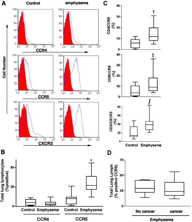
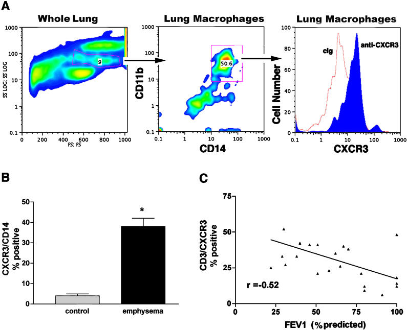
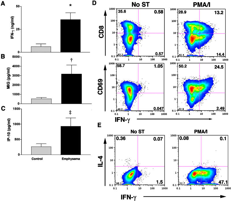
Methods and findings: We studied different subsets of lymphocytes taken from portions of human lungs removed surgically to find out which lymphocytes were the most frequent, which cell-surface markers these lymphocytes expressed, and whether the lymphocytes secreted any specific factors that could be associated with disease. We found that loss of lung function in patients with chronic obstructive pulmonary disease and emphysema was associated with a high percentage of CD4+ and CD8+ T lymphocytes that expressed chemokine receptors CCR5 and CXCR3 (both markers of T helper 1 cells), but not CCR3 or CCR4 (markers of T helper 2 cells). Lung lymphocytes in patients with chronic obstructive pulmonary disease and emphysema secrete more interferon gamma--often associated with T helper 1 cells--and interferon-inducible protein 10 and monokine induced by interferon, both of which bind to CXCR3 and are involved in attracting T helper 1 cells. In response to interferon-inducible protein 10 and monokine induced by interferon, but not interferon gamma, lung macrophages secreted macrophage metalloelastase (matrix metalloproteinase-12), a potent elastin-degrading enzyme that causes tissue destruction and which has been linked to emphysema.

Conclusions: These data suggest that Th1 lymphoctytes in the lungs of people with smoking-related damage drive progression of emphysema through CXCR3 ligands, interferon-inducible protein 10, and monokine induced by interferon.


Conflict of interest statement

Competing Interests: The authors have declared that no competing interests exist.

Figures

Figure 1
Figure 1. Chemokine Receptor Expression on Peripheral Lung Lymphocytes
(A) Single color histograms showing expression of chemokine receptors CCR4, CCR5, and CXCR3 from representative control and emphysema participants. (B) Pooled data from all participants (control, n = 10; emphysema, n = 18) showing percent (median ± SD) of total lung lymphocytes expressing CCR4 and CCR5. (C) Pooled data from same participants showing percent (median ± SD) CCR5 expression on CD4 (top) and CD8 (middle) T cells, and CXCR3 expression on unfractionated T cells (bottom) from the same participant groups. (D) Analysis of total lung lymphocyte chemokine receptor (median ± SD) profiles among participants with emphysema. Participants had either (1) lung volume reduction surgery for emphysema (non-cancer, n = 8) or (2) lung resection for treatment of small peripheral cancer (n = 10). Participants showed similar inflammatory indices as determined by CCR5 expression. In (B) and (C), *, p < 0.001; †, p = 0.01; ‡, p = 0.02; ∫, p = 0.007 (Mann-Whitney test) for the comparison of emphysema and control groups.
Figure 2
Figure 2. Expression of CXCR3 in Lungs of Control and Emphysematous Smoker Individuals
(A) Representative forward and side-scatter characteristics of whole lung cells from a participant with COPD and emphysema. Anti-CD11b PE-conjugated and anti-CD14 FITC-conjugated antibodies detect lung macrophages (middle), and histogram of mean fluorescence intensity showing anti-CXCR3-Cy5 and control antibodies (cIg) detects lung macrophages in the patient with emphysema. (B) Pooled data from control individuals without (n = 5) and with (n = 8) emphysema. Columns are median, bars represent SD. *, p = 0.009 (Mann-Whitney test) for the comparison of emphysema and control participants. (C) Negative association between CXCR3 expression on CD3+ T cells and FEV1 percentage predicted based on an R2 goodness-of-fit statistic of 0.27 (p = 0.0089, r = −0.52, n = 24).
Figure 3
Figure 3. IFN-γ, MIG, and IP-10 Production by Isolated Lung Lymphocytes
(A–C) Lung lymphocytes from control individuals and participants with emphysema were cultured without additional stimulation for 3 or 4 d and assessed for secretion of (A) IFN-γ, (B) MIG, and (C) IP-10 (control, n = 8; emphysema, n = 12). Columns are median, bars represent SD. *, p= 0.007; †, p = 0.01; ‡, p = 0.02 for the comparison of emphysema and control participants. (D) The same cells from a representative ex-smoker individual with emphysema were either left unstimulated (No ST) or treated with PMA/ionomycin (PMA/I) for 24 h and assessed for surface CD8 and CD69 expression and the intracytoplasmic accumulation of IFN-γ by flow cytometry. (E) Production of IL-4 by lung lymphocytes. Lung lymphocytes from a representative ex-smoker individual with emphysema were cultured for 24 h with or without PMA/ionomycin stimulation (PMA/I) and assessed for intracytoplasmic IL-4 and IFN-γ accumulation by flow cytometry.
Figure 4
Figure 4. Regulation of MMP12 by Type 1 Cytokines
(A) CD14+, lymphocyte-depleted lung leukocytes were cultured with and without the indicated amounts of recombinant human IP-10 and IFN-γ, and supernatants were assessed for the presence of MMP12 by Western blotting. (B) Fold increase relative to unstimulated of MMP12 mRNA from lung macrophages stimulated without () and with (+) 500 ng/ml of IP-10 in the presence or absence of a function-blocking antibody to CXCR3 as determined by real-time PCR. (C and D) Lung tissue from a participant with emphysema (C) shows strong immune staining for MMP12 localized to macrophages (arrows), and (D) shows lung tissue from a control participant without emphysema and with undetectable MMP12. The insets show a high-power view of lung macrophages staining positive (C) and negative (D) for MMP12 (×60) *, p = 0.04.

Similar articles

Cited by

References

    1. Barnes PJ. Potential novel therapies for chronic obstructive pulmonary disease. Novartis Found Symp. 2001;234:255–267. - PubMed
    1. Senior RM. Mechanisms of COPD: Conference summary. Chest. 2000;117:320S–323S. - PubMed
    1. Hogg JC. Chronic obstructive pulmonary disease: An overview of pathology and pathogenesis. Novartis Found Symp. 2001;234:4–19. - PubMed
    1. Corry DB, Grunig G, Hadeiba H, Kurup VP, Warnock ML, et al. Requirements for allergen-induced airway hyperreactivity in T and B cell-deficient mice. Mol Med. 1998;4:344–355. - PMC - PubMed
    1. Grunig G, Warnock M, Wakil AE, Venkayya R, Brombacher F, et al. Requirement for IL-13 independently of IL-4 in experimental asthma. Science. 1998;282:2261–2263. - PMC - PubMed

Publication types

MeSH terms