Anatomical study of the paraspinal approach to the lumbar spine
- PMID: 15526219
- PMCID: PMC3489211
- DOI: 10.1007/s00586-004-0802-5
Anatomical study of the paraspinal approach to the lumbar spine
Abstract
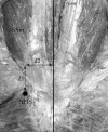
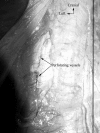
The original description of the paraspinal posterior approach to the lumbar spine was for spinal fusion, especially regarding lumbosacral spondylolisthesis treatment. In spite of the technical details described by Wiltse, exact location of the area where the sacrospinalis muscle has to be split remains somewhat unclear. The goal of this study was to provide topographic landmarks to facilitate this surgical approach. Thirty cadavers were dissected in order to precisely describe the anatomy of the trans-muscular paraspinal approach. The level of the natural cleavage plane between the multifidus and the longissimus part of the sacrospinalis muscle was noted and measurements were done between this level and the midline at the level of the spinous process of L4. A natural cleavage plane between the multifidus and the longissimus part of the sacrospinalis muscle was present in all cases. There was a fibrous separation between the two muscular parts in 55 out of 60 cases. The mean distance between the level of the cleavage plane and the midline was 4 cm (2.4-5.5 cm). In all cases, small arteries and veins were present, precisely at the level of the cleavage plane. We found it possible to easily localize the anatomical cleavage plane between the multifidus part and the longissimus part of the sacrospinalis muscle. First the superficial muscular fascia is opened near the midline, exposing the posterior aspect of the sacrospinalis muscle. Then, the location of the muscular cleft can be found by identifying the perforating vessels leaving the anatomical inter-muscular space.
Figures


Comment in
-
The Michel Benoist and Robert Mulholland yearly European Spine Journal review: a survey of the "surgical and research" articles in the European Spine Journal, 2005.Eur Spine J. 2006 Jan;15(1):8-15. doi: 10.1007/s00586-005-1062-8. Epub 2006 Jan 13. Eur Spine J. 2006. PMID: 16411129 Free PMC article. Review. No abstract available.
References
-
- Dubousset J (1997) Treatment of spondylolysis and spondylolisthesis in children and adolescents. Clin Orthop 77–85 - PubMed
-
- Ray CD (1987) The paralateral approach to decompression for lateral stenosis and far lateral lesions of the lumbar spine. In: Watkins, Collis (eds) Lumbar discectomy and laminectomy. Aspen pp 217–227
-
- Watkins J Bone Joint Surg Am. 1959;41:388. - PubMed
-
- Watkins Clin Orthop. 1964;35:80. - PubMed
-
- Wiltse Clin Orthop. 1964;35:116. - PubMed
Publication types
MeSH terms
LinkOut - more resources
Full Text Sources

