. 2005 Feb 15;386(Pt 1):47-56.
doi: 10.1042/BJ20041054.

Gelsolin binds to polyphosphoinositide-free lipid vesicles and simultaneously to actin microfilaments

Affiliations

Gelsolin binds to polyphosphoinositide-free lipid vesicles and simultaneously to actin microfilaments

Jocelyn Méré et al. Biochem J. .

Abstract

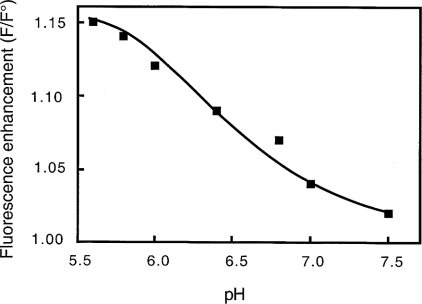
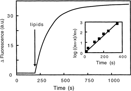
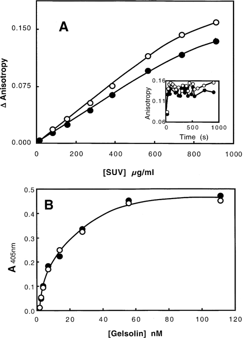
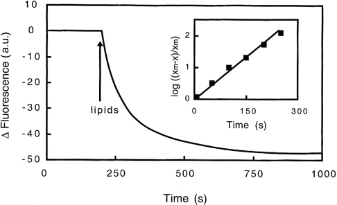
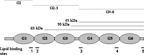
Gelsolin is a calcium-, pH- and lipid-dependent actin filament severing/capping protein whose main function is to regulate the assembly state of the actin cytoskeleton. Gelsolin is associated with membranes in cells, and it is generally assumed that this interaction is mediated by PPIs (polyphosphoinositides), since an interaction with these lipids has been characterized in vitro. We demonstrate that non-PPI lipids also bind gelsolin, especially at low pH. The data suggest further that gelsolin becomes partially buried in the lipid bilayer under mildly acidic conditions, in a manner that is not dependent of the presence of PPIs. Our data also suggest that lipid binding involves a number of sites that are spread throughout the gelsolin molecule. Linker regions between gelsolin domains have been implicated by other work, notably the linker between G1 and G2 (gelsolin domains 1 and 2 respectively), and we postulate that the linker region between the N-terminal and C-terminal halves of gelsolin (between G3 and G4) is also involved in the interaction with lipids. This region is compatible with other studies in which additional binding sites have been located within G4-6. The lipid-gelsolin interactions reported in the present paper are not calcium-dependent, and are likely to involve significant conformational changes to the gelsolin molecule, as the chymotryptic digest pattern is altered by the presence of lipids under our conditions. We also report that vesicle-bound gelsolin is capable of binding to actin filaments, presumably through barbed end capping. Gelsolin bound to vesicles can nucleate actin assembly, but is less active in severing microfilaments.


Figures

Figure 1
Figure 1. Effects of pH on the interaction of gelsolin with PG/PC SUVs
Interaction of gelsolin with SUVs (0.4 mg/ml) (25% PG+75% PC) was monitored by fluorescence. Enhancement (F/F°) in the intensity of the fluorescence emission spectra of gelsolin tryptophan residues were recorded at various pH between 5.6 and 7.5 in 0.15 M NaCl, 1 mM EGTA and 0.05 M Mes or Tris/HCl buffers. Excitation was fixed at 280 nm and emission was recorded at 340 nm.
Figure 2
Figure 2. Kinetics of the interaction of gelsolin with PG/PC SUVs at pH 5.6 monitored by fluorescence
SUVs (25% PG+75% PC, 0.4 mg/ml) were added to gelsolin in 0.15 M NaCl, 5 mM EGTA and 0.05 M acetate buffer, pH 5.6, and changes in fluorescence intensity of gelsolin tryptophan residues at 340 nm were recorded over time. Inset, graphical determination of the constant rate related to the fluorescence enhancement induced by gelsolin interaction with PG/PC SUVs, log [(xm−x)/xm] is plotted against time, where xm is the maximum fluorescence change observed and x is the fluorescence change at time t. a.u., arbitrary units.
Figure 3
Figure 3. Binding of gelsolin to PG/PC SUVs
(A) Interaction of gelsolin monitored by tryptophan fluorescence anisotropy. Experiments were carried out in 0.15 M NaCl, 5 mM EGTA and 0.05 M acetate buffer, pH 5.6 (●), or 0.15 M NaCl, 1 mM EGTA and 0.05 M Tris/HCl buffer, pH 7.0 (○). Anisotropy change is plotted against PG/PC SUV concentration. Inset, PG/PC SUVs (0.4 mg/ml) were added to gelsolin at zero time, and anisotropy was recorded over time at pH 5.6 (●) or pH 7.0 (○). (B) Interaction of gelsolin with coated phospholipids (0.04 mg/ml) (25% PG+75% PC). Biotinylated gelsolin was added at various concentrations (0–110 nM) at pH 5.6 (●) or pH 7.0 (○) in the same buffers, and the binding was monitored at 405 nm.
Figure 4
Figure 4. Kinetics of the interaction of gelsolin with Br-PC/PG SUVs at pH 5.6 monitored by fluorescence
SUVs (25% PG, 37.5% Br-PC and 37.5% PC) at 0.4 mg/ml were added to gelsolin in 0.15 M NaCl, 5 mM EGTA and 0.05 M acetate buffer, pH 5.6, and fluorescence quenching of gelsolin tryptophan residues was recorded over time. Inset, graphical determination of the constant rate related to the fluorescence quenching induced by gelsolin interaction with Br-PC/PG SUVs, log [(xm−x)/xm] is plotted against time, where xm is the maximum fluorescence change observed and x is the fluorescence change at time t. a.u., arbitrary units.
Figure 5
Figure 5. Quenching of tryptophan fluorescence of gelsolin by acrylamide
Stern–Volmer plot for the quenching of gelsolin by acrylamide in 0.15 M NaCl, 5 mM EGTA and 0.05 M acetate buffer, pH 5.6 (○) or 0.15 M NaCl, 1 mM EGTA and 0.05 M Tris/HCl buffer, pH 7.0 (□). The same experiments were also performed in the presence of PG/PC SUVs (0.4 mg/ml) (25% PG+75% PC) at pH 5.6 (●) or pH 7.0 (■). F°/F was determined as described in the Materials and methods section. The excitation wavelength was set at 280 nm.
Figure 6
Figure 6. Interaction of gelsolin with phospholipids measured by FRET
Tryptophan emission spectra of gelsolin–PG/PC SUV complexes were performed in 5 mM EGTA and 0.05 M acetate buffer, pH 5.6, in the absence (—) and in the presence of 4 μM DPH (-·-·-). Gelsolin was used at 0.4 μM and PG/PC SUVs (25% PG+75% PC) were at 0.4 mg/ml. Excitation was at 280 nm and emission was recorded between 300 and 500 nm. The emission spectrum between 400 and 500 nm of the tertiary complex resulting from the excitation of DPH fluorophore at 350 nm is also included (-··-··-··-, secondary y-axis). a.u., arbitrary units.
Figure 7
Figure 7. Effect of PG/PC SUVs on susceptibility of gelsolin to chymotryptic digestion at pH 7.0
Digestion of gelsolin in 0.15 M NaCl, 1 mM CaCl2 and 0.05 M Tris/HCl buffer, pH 7.0, by chymotrypsin (chymotrypsin/gelsolin ratio, 1:300, w/w) for various times (3, 6, 10 and 15 min) in the absence (lanes 2–5 respectively) or in the presence of 0.4 mg/ml PG/PC SUVs (25% PG+75% PC), (lanes 6–9 respectively). Gelsolin at zero time is in lane 1. Molecular-mass markers are in lane M; standards are phosphorylase B (97.4 kDa), BSA (66.2 kDa), ovalbumin (45 kDa), carbonic anhydrase (31 kDa), soybean trypsin inhibitor (21.5 kDa) and lysozyme (14.5 kDa). Samples were separated by SDS/15% (w/v) PAGE.
Figure 8
Figure 8. Effects of PG/PC SUVs on the susceptibility of gelsolin to chymotryptic digestion at pH 6
(A) Digestion of gelsolin in 0.1 M Mes buffer, pH 6.0, by chymotrypsin (chymotrypsin/gelsolin ratio, 1:200, w/w) for various times (3, 6, 10 and 15 min) in the absence (lanes 2–5 respectively) or in the presence of 0.4 mg/ml PG/PC SUVs (25% PG+75% PC) (lanes 6–9 respectively). Gelsolin at zero time is in lane 1. Molecular-mass markers are in lane M and are as in Figure 7. Samples were separated by SDS/12.5% PAGE. (B) Kinetics of chymotryptic digestion. Bands in the electrophoretic gel presented above were integrated and the intensity plotted against time. Results are for 90 kDa (gelsolin) compound with (△) or without PG/PC SUVs (□), and 63 kDa fragment with (×) or without PG/PC SUVs (◇). a.u., absorbance units.
Figure 9
Figure 9. Tryptophan fluorescence of gelsolin (1), G1–3 (2) and G4–6 (3) in the presence of PG/PC SUVs and Br-PC/PG SUVs at pH 5.6
Figure 10
Figure 10. Effect of PG/PC SUVs on the activation of gelsolin nucleating activity
Labelled 2.3 μM G-actin was added to 2 mM MgCl2 and 0.1 M KCl, 20 μM ATP and 20 mM Mes buffer at pH 5.8 (A) or 2 mM MgCl2 and 0.1 M KCl, 20 μM ATP, 0.5 mM CaCl2 and 20 mM Tris/HCl buffer at pH 7.5 (B) in the presence (circles) or in the absence (squares) of 26 nM gelsolin, as described in the Materials and methods section. The increase in fluorescence in the presence (open symbols) or in the absence (closed symbols) of PG/PC SUVs (25% PG+75% PC) at 0.2 mg/ml was plotted against time. a.u., arbitrary units.
Figure 11
Figure 11. Confocal image of actin filaments in association with PG/PC SUVs and gelsolin
G-actin (3 μM) was added to 2 mM MgCl2 and 0.1 M KCl, 20 μM ATP and 20 mM Mes buffer at pH 5.8 or 2 mM MgCl2 and 0.1 M KCl, 20 μM ATP, 0.5 mM CaCl2 and 20 mM Tris/HCl buffer at pH 7.5 in the presence of PG/PC SUVs at 0.2 mg/ml (25% PG+75% PC) containing 2% NBD-labelled PC. Actin filaments were stained with rhodamine-labelled phalloidin before visualization. (A), (a), (B) and (b) show experiments performed at pH 5.8 in the presence or absence of 52 nM gelsolin respectively. (C), (c), (D) and (d) show experiments performed at pH 7.5 in the presence or absence of 52 nM gelsolin respectively. Scale bar, 2 μm.
Figure 12
Figure 12. Effects of PG/PC SUVs on the severing activity of gelsolin
(A) Labelled 25 μM F-actin (pre-capped by 1:1000 molar ratio of gelsolin) was diluted to 480 nM in either 2 mM MgCl2 and 0.1 M KCl, 20 μM ATP and 20 mM Mes buffer at pH 5.8 (left-hand panel) or 2 mM MgCl2 and 0.1 M KCl, 20 μM ATP, 0.5 mM CaCl2, 20 mM Tris/HCl buffer at pH 7.5 (right-hand panel) in the presence of 52 nM gelsolin (squares) or in the absence of gelsolin (circles). Fluorescence at 386 nm (a.u., arbitrary units) was reported over time in the absence (open symbols) and in the presence (closed symbols) of PG/PC SUVs (25% PG+75% PC) at 0.2 mg/ml. (B) Rates of depolymerization were measured from experiments conducted as in (A). Left-hand panel, pH 5.8 buffer; right-hand panel, pH 7.5 buffer, with 52 nM (lanes 1 and 2) or 26 nM (lanes 3 and 4) gelsolin. The rate of actin depolymerization (a.u.) was reported in the absence (lanes 1 and 3) and in the presence (lanes 2 and 4) of PG/PC SUVs (25% PG+75% PC) at 0.2 mg/ml.
Figure 13
Figure 13. Domain structure of gelsolin showing proteolytic products at pH 7.0 (broken lines) and pH 6.0 (solid lines, 45, 50 and 63 kDa products) in the presence of PG/PC SUVs
Lipid-binding regions are also shown in the lower part of the diagram: 1 and 2, the well-documented PtdIns(4,5)P2-binding sites 135–149 and 150–169; 3, a possible lipid-binding site suggested by the present study; 4 and 5, other sites in G4–6.

Similar articles

Cited by

References

    1. dos Remedios C. G., Chhabra D., Kekic M., Dedova I. V., Tsubakihara M., Berry D. A., Noseworthy N. J. Actin binding proteins: regulation of cytoskeletal microfilaments. Physiol. Rev. 2003;83:433–473. - PubMed
    1. Isenberg G., Goldmann W. H. Actin-binding protein–lipid interaction. In: Pryme H., editor. The Cytoskeleton, vol. 1. Greenwich, CT/London: JAI Press Inc.; 1995. pp. 169–204.
    1. Irvine R. F., Schell M. J. Back in the water: the return of the inositol phosphates. Nat. Rev. Mol. Cell Biol. 2001;2:327–338. - PubMed
    1. Dowler S., Currie R. A., Campbell D. G., Deak M., Kular G., Downes C. P., Alessi D. R. Identification of pleckstrin-homology-domain-containing proteins with novel phosphoinositide-binding specificities. Biochem. J. 2000;351:19–31. - PMC - PubMed
    1. Ellson C. D., Andrews S., Stephens L. R., Hawkins P. T. The PX domain: a new phosphoinositide-binding module. J. Cell Sci. 2002;115:1099–1105. - PubMed

Publication types

MeSH terms