. 2005 Feb 1;562(Pt 3):655-72.
doi: 10.1113/jphysiol.2004.073338. Epub 2004 Nov 4.

Localization and function of the Kv3.1b subunit in the rat medulla oblongata: focus on the nucleus tractus solitarii

Affiliations

Localization and function of the Kv3.1b subunit in the rat medulla oblongata: focus on the nucleus tractus solitarii

Mark L Dallas et al. J Physiol. .

Abstract

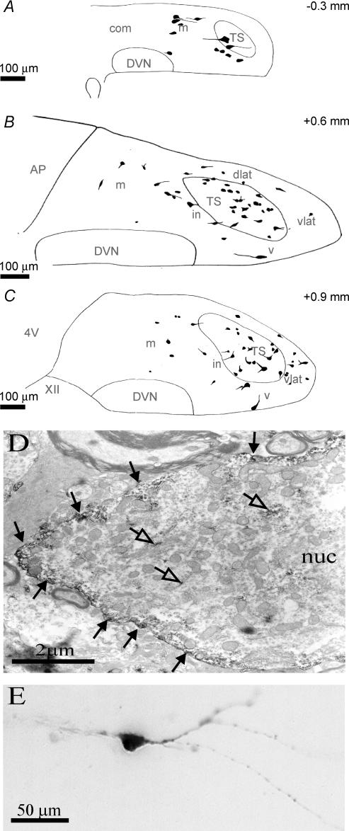
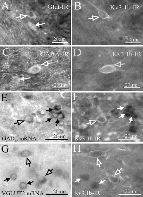
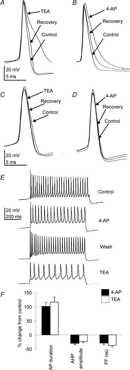
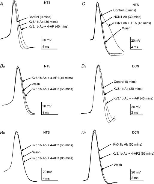
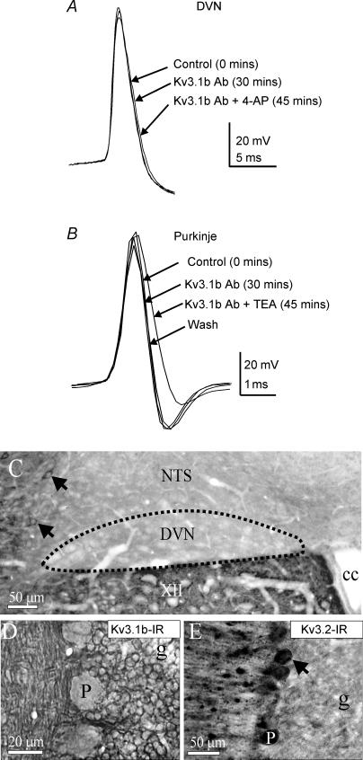
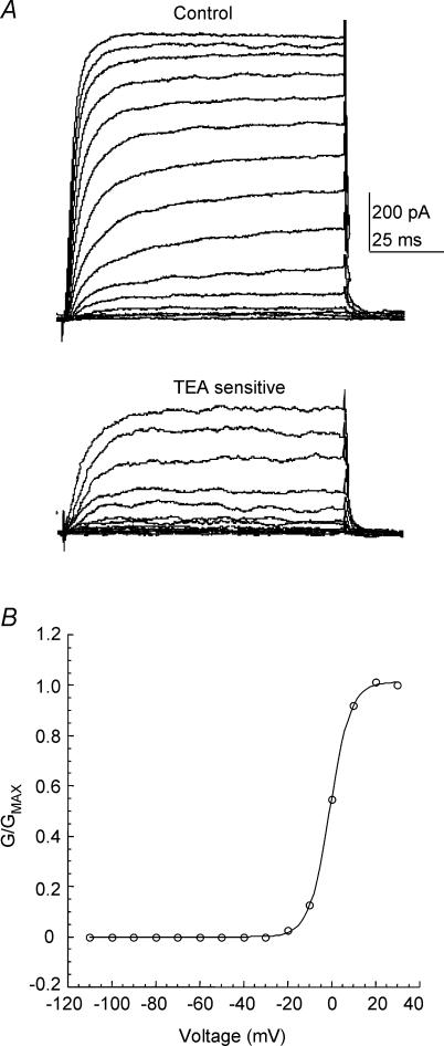
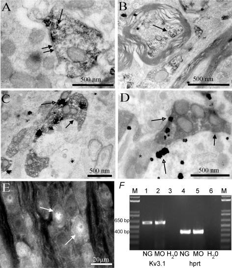
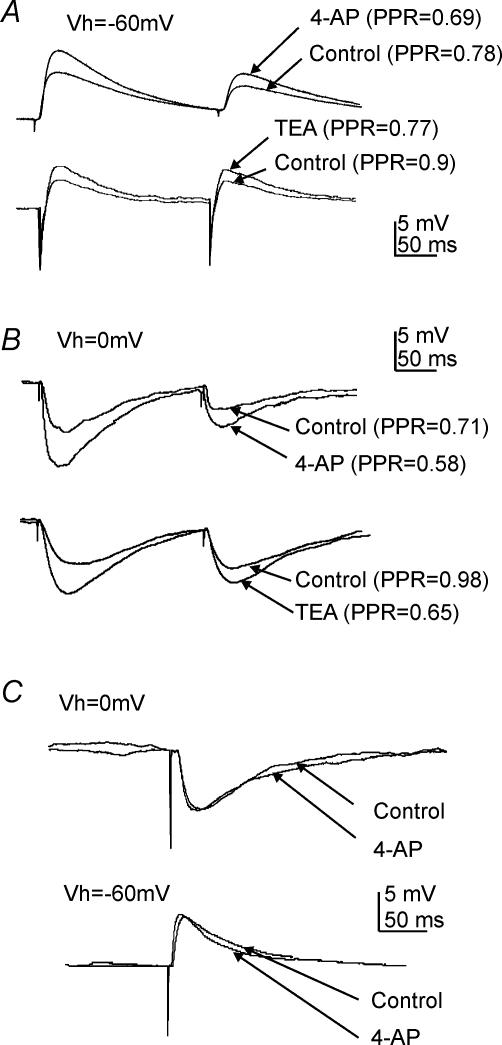
The voltage-gated potassium channel subunit Kv3.1 confers fast firing characteristics to neurones. Kv3.1b subunit immunoreactivity (Kv3.1b-IR) was widespread throughout the medulla oblongata, with labelled neurones in the gracile, cuneate and spinal trigeminal nuclei. In the nucleus of the solitary tract (NTS), Kv3.1b-IR neurones were predominantly located close to the tractus solitarius (TS) and could be GABAergic or glutamatergic. Ultrastructurally, Kv3.1b-IR was detected in NTS terminals, some of which were vagal afferents. Whole-cell current-clamp recordings from neurones near the TS revealed electrophysiological characteristics consistent with the presence of Kv3.1b subunits: short duration action potentials (4.2 +/- 1.4 ms) and high firing frequencies (68.9 +/- 5.3 Hz), both sensitive to application of TEA (0.5 mm) and 4-aminopyridine (4-AP; 30 mum). Intracellular dialysis of an anti-Kv3.1b antibody mimicked and occluded the effects of TEA and 4-AP in NTS and dorsal column nuclei neurones, but not in dorsal vagal nucleus or cerebellar Purkinje cells (which express other Kv3 subunits, but not Kv3.1b). Voltage-clamp recordings from outside-out patches from NTS neurones revealed an outward K(+) current with the basic characteristics of that carried by Kv3 channels. In NTS neurones, electrical stimulation of the TS evoked EPSPs and IPSPs, and TEA and 4-AP increased the average amplitude and decreased the paired pulse ratio, consistent with a presynaptic site of action. Synaptic inputs evoked by stimulation of a region lacking Kv3.1b-IR neurones were not affected, correlating the presence of Kv3.1b in the TS with the pharmacological effects.


Figures

Figure 1
Figure 1. Kv3.1b immunoreactivity is widely distributed throughout the medulla oblongata
A, a low-power montage of Kv3.1b subunit immunoreactivity (Kv3.1b-IR) in the medulla oblongata (detected using a Cy3-conjugated secondary antibody; left-hand side) indicating that Kv3.1b-IR is present in many regions of the medulla oblongata. The right-hand side is a schematic diagram to aid orientation of the sections by illustrating the location of various brain regions according to the Paxinos & Watson, 1986 rat brain atlas. Higher magnifications of Kv3.1b immunoreactivity in selected areas are illustrated in BG. B, numerous neurones were labelled in the cuneate nucleus. C, all neurones in the gracile nucleus contained Kv3.1b-IR. D, a large Kv3.1b-IR neurone in the dorsal medullary reticular nucleus. E, example of a rare labelled neurone within the nucleus ambiguus (filled arrow). Also shown is a vagal preganglionic neurone (open arrow) which was identified by retrograde tracing with fluorogold (not shown). Typically, the vagal preganglionic neurone is apposed by Kv3.1b-immunoreactive structures, but does not contain the channel itself. F, Kv3.1b-IR neurones from the spinal trigeminal nucleus. G, a large presumptive motorneurone in the hypoglossal nucleus (XII) that is apposed by Kv3.1b-IR structures, but does not appear to contain the subunit within its membrane. Punctate Kv3.1b immunoreactivity is also present throughout the neuropil. AP, area postrema; cu, cuneate fasiculus; Cu, cuneate nucleus; DVN, dorsal vagal motor nucleus; Gr gracile nucleus; Lrt, lateral reticular nucleus; MdD, dorsal medullary reticular nucleus; MdV, ventral medullary reticular nucleus; NA, nucleus ambiguus; NTS, nucleus tractus solitarii; py, pyramidal tract; PMn, paramedian reticular nucleus; sp5, spinal trigeminal tract; Sp5, spinal trigeminal nucleus.
Figure 2
Figure 2. Kv3.1b-immunoreactive neurones in the NTS are located predominantly in the vicinity of the solitary tract
A–C, camera lucida drawings of Kv3.1b-immunoreactive neurones superimposed on sketches of NTS sections from three different rostrocaudal levels with respect to the obex (levels in top right hand corner). A, at caudal levels (at the level of the central canal) a few Kv3.1b-IR neurones were observed in the medial (m) subdivision of the NTS and within the tractus solitarius (TS). B, at the level of the area postrema (AP), a greater number of Kv3.1b-IR cells were present. A few Kv3.1b-IR neurones were located in the medial subnucleus, but a greater proportion were located in the dorsolateral (dLat), interstitial (in), ventral (v) and ventrolateral (vlat) subdivisions and within the TS. C, at the level of the fourth ventricle (4V), a similar distribution of Kv3.1b-IR neurones was observed to that at the level of the area postrema. Again, a few Kv3.1b-IR neurones were located in the medial subnucleus, but a greater proportion were located in the dLat, in, v and vlat subdivisions and within the TS. D, electron micrograph of a Kv3.1b-IR neurone in the NTS visualized with diaminobenzidine. The Kv3.1b reaction product is apposed to the membrane (filled arrows), suggesting that the channel is inserted into the membrane. Immunoreactivity is also sparsely distributed throughout the cytoplasm (open arrows); nuc, nucleus. E, NTS neurone filled with Lucifer yellow which was sensitive to TEA and 4-AP. The neurone was located in the ventral region of the tractus and the projections run in the mediolateral axis.
Figure 3
Figure 3. Kv3.1b immunoreactivity is present in both GABA- and glutamate-containing neurones in the NTS
A, two glutamate-immunoreactive neurones in the vicinity of the TS (arrows, visualized by Alexa488). The open arrow points to one of the cells that is also Kv3.1b immunoreactive, as shown in B. B, same area as A, but viewed through a Cy3 filter set and showing a Kv3.1b-IR neurone (open arrow) that is also glutamate-IR as depicted in panel A. C, two GABA-IR neurones in the vicinity of the TS (arrows, visualized by Alexa488). The open arrow highlights one of the cells that is also Kv3.1b-IR, as indicated in D. D, same area as C, but viewed through a Cy3 filter set, and showing a Kv3.1b-IR neurone (arrow) that is also GABA-IR, as depicted in C. E, neurones in the NTS expressing mRNA encoding the GAD65 isoform of the GABA-synthesizing enzyme glutamic acid decarboxylase (arrows). The open arrow highlights a labelled neurone that is also Kv3.1b-IR, as shown in F. F, visualization of the same region as in E with a Cy3 filter set to reveal Kv3.1b-IR indicates that one of the GAD65-expressing neurones is also Kv3.1b-IR (open arrow). The other GAD65-expressing neurones do not contain Kv3.1b-IR (filled arrows). G, neurones in the NTS expressing the mRNA for the vesicular glutamate transporter VGLUT2 (arrows). Two of these labelled neurones (open arrows) are also Kv3.1b-IR, as shown in H. Neurones indicated with filled arrows are not Kv3.1b-IR. H, visualizing Kv3.1b immunoreactivity in the same region as G reveals that two of the VGLUT2 expressing neurones are also Kv3.1b-IR (open arrows). Other VGLUT2-expressing neurones are not Kv3.1b-IR (filled arrows).
Figure 4
Figure 4. Low concentrations of TEA and 4-AP prolong repolarization of action potentials in NTS neurones
A, application of 0.5 mm TEA to an NTS neurone increased the action potential duration by prolonging the repolarization phase, an effect that was reversed on washout. B, application of 30 μm 4-AP to the same NTS neurone as in A similarly increased the action potential duration, and the effect was reversed on washout. These data were collected 1.5 h into the recording. Note the control action potential did not change from that in A, recorded at 25 min. C and D, examples of DVN neurones where 0.5 mm TEA (C) and 30 μm 4-AP (D) used at the same concentrations as on NTS neurones had no significant effect on the action potential duration. E, application of 4-AP to an NTS neurone reduced the number of action potentials fired in response to a +60 pA current pulse by 33%, an effect reversed on washout. Application of TEA to the same neurone also reduced the number of action potentials elicited (59% reduction). F, pooled data showing the effects of 4-AP and TEA on the action potential duration, afterhyperpolarization (AHP) amplitude and the steady-state firing frequency in NTS neurones as a percentage change from the control state.
Figure 5
Figure 5. Intracellular application of an anti-Kv3.1b antibody, but not anti-HCN1, prolongs the action potential duration in neurones in the NTS
A, when an anti-Kv3.1b antibody (0.3 μg ml−1) was present in the patch pipette, the action potential duration of the recorded NTS neurone increased gradually over time. This duration was further increased by subsequent application of 30 μm 4-AP. Ba, The same neurone as in A at a later time point. The effect of 4-AP (4-AP1) was partially reversed on washout and further application of 4-AP (4-AP2) prolonged the action potential duration as before. Bb, further application of 4-AP (4-AP3) at 85 min did not increase the action potential duration from that of the previous application (4-AP2), suggesting that the antibody had completely occluded the effects of 4-AP. C, example of an NTS neurone in which the hyperpolarization-activated cyclic nucleotide-gated channel 1 subunit (HCN1) antibody was added to the intracellular solution at the same concentration as the Kv3.1b antibody (0.3 μg ml−1). The antibody alone had no effect on the action potential waveform, and did not alter the sensitivity to TEA (100 μm). Da, example of a DCN neurone in which intracellular application of the Kv3.1b antibody led to a progressive increase in the action potential duration, and this was further prolonged by bath application of 4-AP (30 μm). Db, the effect of 4-AP was occluded, since at t = 50 min, 4-AP (4-AP2) had no further effect on the action potential duration.
Figure 6
Figure 6. Intracellular application of the Kv3.1b antibody does not affect neurones which do not contain Kv3.1b, but which do contain other Kv3 subunits
A, in this DVN neurone, intracellular application of the Kv3.1b antibody and 30 μm 4-AP did not affect the action potential waveform. B, example of a cerebellar Purkinje neurone in which application of the Kv3.1b antibody did not affect the action potential shape, and did not interfere with sensitivity of the neurone to TEA (100 μm), presumably acting on the other Kv3 subunits in these cells. C, low-power image showing that Kv3.1b-IR (detected using DAB) is absent from the DVN. Labelled Kv3.1b-IR cells can clearly be seen in the interstitial region of the NTS (arrows), and punctate Kv3.1b-IR can also be observed in the hypoglossal nucleus (XII) in the same section; cc, central canal. D, high-power light microscope image showing Kv3.1b-IR is absent from cerebellar Purkinje cell somata. E, low-power light micrograph showing Kv3.2-IR is present in cerebellar Purkinje cell soma.
Figure 7
Figure 7. TEA-sensitive K+ currents in α-DTX-sensitive outside-out patches from NTS neurones
A, upper panel, representative example of voltage-dependent K+ currents recorded from an outside-out patch excised from an NTS neurone in response to 100 ms depolarizing voltage steps (in 10 mV increments) from a holding potential of −110 mV. Lower panel, offline digital subtraction of the current in the presence of TEA (100 μm; not shown) from the control current (A) reveals a TEA-sensitive component. B, representative example of the steady-state activation curve for the TEA-sensitive current. Data points were obtained by calculating the conductance of the TEA-sensitive component and plotting normalized values against the voltage step. A single, first-order Boltzmann function (see Methods for equation) is superimposed on the data points. Notice the threshold of around −20 mV, and the depolarized V½ value.
Figure 8
Figure 8. The Kv3.1b subunit is present in presynaptic terminals in the NTS, some of which are vagal afferent fibre terminals
A, electron micrograph of a Kv3.1b-immunoreactive terminal in the NTS identified by diaminobenzidine reaction product (arrow) that forms a synaptic contact (double arrows) with an unlabelled dendrite. B, a myelinated axon in the TS contains Kv3.1b immunoreactivity (arrow). C, a vagal afferent fibre terminal, identified by the presence of diaminobenzidine reaction product following detection of anterograde tracer injected into the nodose ganglion, makes synaptic contacts (double arrows) in the NTS. The terminal also contains silver-intensified gold (open arrow) indicating that it is Kv3.1b immunoreactive. D, an anterogradely labelled, Kv3.1b containing vagal afferent fibre terminal in the NTS forms a synaptic contact (filled arrow) with a Kv3.1b-immunoreactive neurone identified by silver-intensified gold particles (open arrows). An unlabelled terminal (asterisk) also innervates the same neurone. E, Kv3.1b immunoreactivity detected using a Cy3-conjugated secondary antibody in the nodose ganglion is present in the cytoplasm of some neurones, but absent from their membranes. The image has been transformed to greyscale and inverted. F, RT-PCR indicates that Kv3.1 is expressed in the nodose ganglion (NG; lane 1) and medulla oblongata (MO; lane 2). As a positive control for the PCR reaction, cDNA encoding the housekeeping gene hprt was amplified from RNA extracted from the NG (lane 4) and MO (lane 5). There were no amplified products detected when using water as a template (lanes 3 and 6). M, scale ladder.
Figure 9
Figure 9. Pathway-specific modulation of neurotransmitter release by 4-AP and TEA in the NTS
A, paired stimulation of the solitary-tract-elicited EPSPs in an NTS neurone with a paired-pulse ratio (PPR) of 0.78. Application of 30 μm 4-AP (upper trace) increased the amplitude of both the first and second EPSPs, but significantly decreased the PPR to 0.69. Application of 0.5 mm TEA had a similar effect (lower trace). B, IPSPs in an NTS neurone evoked by paired-pulse stimulation of the solitary tract in the presence of kynurenic acid, and at a holding potential of 0 mV. Applications of both 4-AP (upper trace) and TEA (lower trace) increased IPSP amplitude and decreased the PPR. C, stimulation of the reticular formation in a region where Kv3.1b-immunoreactive neurones were not detected elicited EPSPs (upper trace) and IPSPs (lower trace) in NTS neurones on which 4-AP had no effect. All traces are averages of ten single sweeps.

Similar articles

Cited by

References

    1. Archer SL, Souil E, Dinh-Xuan AT, Schremmer B, Mercier JC, El Yaagoubi A, Nguyen-Huu L, Reeve HL, Hampl V. Molecular identification of the role of voltage-gated K+ channels Kv1.5 and Kv2.1 in hypoxic pulmonary vasoconstriction, and control of resting membrane potential in rat pulmonary artery myocytes. J Clin Invest. 1998;101:2319–2330. - PMC - PubMed
    1. Atkinson L, Deuchars J. Distribution of Kv3.1b immunoreactivity in the dorsomedial medulla of the rat. Proc Physiol Soc. 2000;526.P:171.
    1. Baranauskas G, Tkatch T, Nagata K, Yeh JZ, Surmeier DJ. Kv3.4 subunits enhance the repolarizing efficiency of Kv3.1 channels in fast-spiking neurons. Nat Neurosci. 2003;6:258–266. - PubMed
    1. Barraco R, El-Ridi M, Ergene E, Parizon M, Bradley D. An atlas of the rat subpostremal nucleus tractus solitarius. Brain Res Bull. 1992;29:703. - PubMed
    1. Berger AJ. Dorsal respiratory group neurons in the medulla of cat: spinal projections, responses to lung inflation and superior laryngeal nerve stimulation. Brain Res. 1977;135:231–254. - PubMed

Publication types

Substances

LinkOut - more resources