. 2005 Jan 15;562(Pt 2):569-82.
doi: 10.1113/jphysiol.2004.071381. Epub 2004 Nov 4.

Neuronal mechanisms mediating the variability of somatosensory evoked potentials during sleep oscillations in cats

Affiliations

Neuronal mechanisms mediating the variability of somatosensory evoked potentials during sleep oscillations in cats

Mario Rosanova et al. J Physiol. .

Abstract

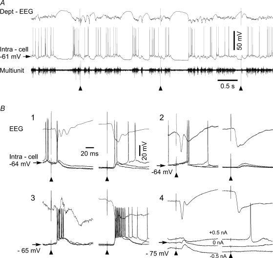
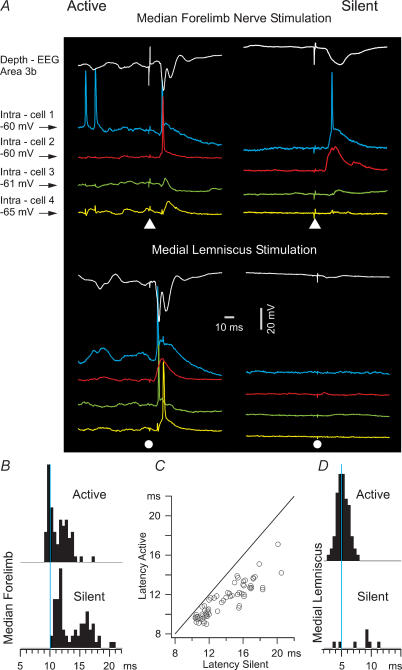
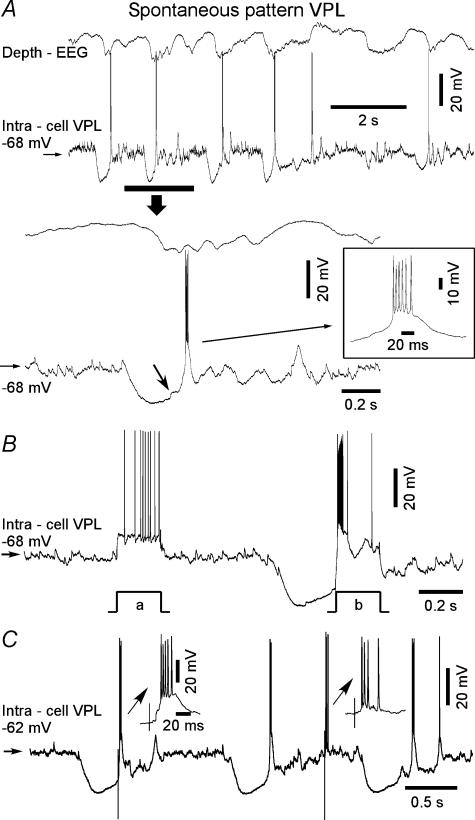
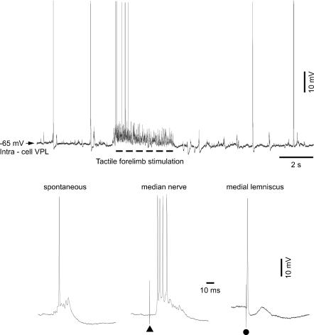
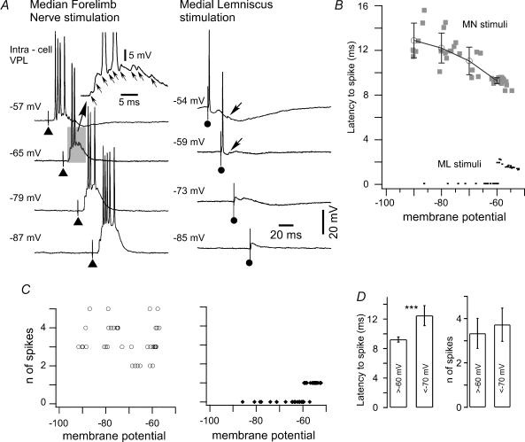
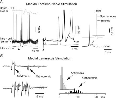
The slow oscillation (SO) generated within the corticothalamic system is composed of active and silent states. The studies of response variability during active versus silent network states within thalamocortical system of human and animals provided inconsistent results. To investigate this inconsistency, we used electrophysiological recordings from the main structures of the somatosensory system in anaesthetized cats. Stimulation of the median nerve (MN) elicited cortical responses during all phases of SO. Cortical responses to stimulation of the medial lemniscus (ML) were virtually absent during silent periods. At the ventral-posterior lateral (VPL) level, ML stimuli elicited either EPSPs in isolation or EPSPs crowned by spikes, as a function of membrane potential. Response to MN stimuli elicited compound synaptic responses and spiked at any physiological level of membrane potential. The responses of dorsal column nuclei neurones to MN stimuli were of similar latency, but the latencies of antidromic responses to ML stimuli were variable. Thus, the variable conductance velocity of ascending prethalamic axons was the most likely cause of the barrages of synaptic events in VPL neurones mediating their firing at different level of the membrane potential. We conclude that the preserved ability of the somatosensory system to transmit the peripheral stimuli to the cerebral cortex during all the phases of sleep slow oscillation is based on the functional properties of the medial lemniscus and on the intrinsic properties of the thalamocortical cells. However the reduced firing ability of the cortical neurones during the silent state may contribute to impair sensory processing during sleep.


Figures

Figure 1
Figure 1. Modulation of cortical somatosensory evoked potential and intracellular activities during slow oscillation
All recordings were obtained from the focus of responses to the electrical stimulation of the right forelimb median nerve in the left somatosensory cortex. A, an example of the focal field, intracellular and multiunit recordings during a session of peripheral stimulation (▴). The first and the second stimuli were applied during active network states and the third stimulus was delivered during the silent network state. B, averaged field potential responses and superimposition of three intracellular single traces selected during active (left side) and silent (right side) network states. Note the multiple components of field responses during active cortical states and their modification during silent states. 1, 2 and 3 are the three types of initial excitatory responses; 4 is the initial inhibitory response. Note the increased latency of excitatory responses that occurred during EEG depth-positive wave and the absence of inhibitory response during the silent network state.
Figure 2
Figure 2. Differences in cortical responses to forelimb versus medial lemniscus stimulation during different phases of SO
A, simultaneous field and quadruple intracellular recording during responses to forelimb median nerve and medial lemniscus stimuli. Forelimb stimulation, ▴; medial lemniscus stimulation, •. White trace: focal field potential; blue, red green and yellow: four different neurones recorded simultaneously. Left panels, responses during active network states, right panels, responses during silent network states. B, histograms of minimal response latency of the 73 neurones that responded to median forelimb nerve during active and silent network states. C, scatter plot for the response latency of cortical neurones during active versus silent states. D, histograms of minimal response latency of the 73 neurones that responded to medial lemniscus stimuli during active and silent network states.
Figure 3
Figure 3. Spontaneous activity and response patterns of the thalamocortical neurone located in the VPL nucleus during slow oscillation
A, an example of simultaneous recordings of cortical field potential from the somatosensory cortex and intracellular recordings from a VPL neurone. The period indicated by the horizontal bar and arrow is expanded in the middle panel. The inset shows a typical spontaneous low-threshold spike burst. Note that the thalamocortical neurone was hyperpolarized during depth-positive EEG waves, displayed a rebound spike burst after the onset of cortical activities as estimated by EEG depth-negativity, and later displayed a brief sequence of spindle-related short IPSPs. B, depolarizing current pulses of 0.5 nA elicited burst firing of the neurone during hyperpolarizing phases of slow oscillation and the same current pulse during elicited tonic firing depolarizing phases of slow oscillation. C, medial nerve stimuli applied during both silent and active states elicit excitation and firing of VPL neurone.
Figure 4
Figure 4. Response of VPL thalamocortical neurone to forelimb versus medial lemniscus stimulation
Upper panel, a period of intracellular activity recorded from a VPL neurone of the thalamus. The thick broken line indicates the time segment during which slight pressure was applied to the skin in the receptive field (distal forelimb) of the TC neurone. Lower panel, from left to right: Spontaneous complex synaptic events during resting membrane potential and responses to median nerve (▴) and medial lemniscus stimuli (•). Note the presence of multiple synaptic events and high-frequency spike trains elicited by forelimb stimulation and a single EPSP leading to spikes elicited by medial lemniscus stimulation.
Figure 5
Figure 5. Voltage dependency of responses of a VPL neurone to forelimb median nerve and medial lemniscus stimuli
A, responses of a VPL neurone to MN (left) and ML (right) stimulation. In order to study the voltage dependency of responses, de- and hyperpolarizing steady DC current was injected intracellularly. At depolarized voltages the forelimb stimulation elicited high-frequency spike trains, whereas at more hyperpolarized voltages the summated multiple synaptic potentials triggered a low-threshold spike burst. Small arrows in the inset point to individual EPSPs contributing to a summated response. Note the increased latency of the first spike at more hyperpolarized voltages. In contrast, medial lemniscus stimulation at hyperpolarized voltages elicited single EPSPs that were crowned with spikes at more depolarized voltages. A small arrow points to the second EPSPs that were observed in some traces. B, the dependency of the first spike latency on the membrane potential. The average values for responses to MN stimuli were obtained in voltage windows from −95 mV to −85 mV, from −85 mV to −75 mV, from −75 mV to −65 mV and from −65 mV to −55 mV. There were no spikes elicited by ML stimuli (indicated as 0 ms on y axis) at voltages below −60 mV. C, the dependency of number of spikes on the membrane potential for forelimb (left) and medial lemniscus (right) electrical stimuli from the neurone shown in A. Note that at any given level of membrane potential, forelimb stimulation was able to trigger a high-frequency sequence of spikes (from two to five). In contrast, medial lemniscus stimulation revealed a binary behaviour characterized by the absence of spikes at hyperpolarized membrane potentials (until −60 mV) and one spike at more depolarized levels of membrane potential. D, mean values (n = 7 neurones) of first spike latency and the number of spikes at depolarized voltages of more than −60 mV and at hyperpolarized voltages of more than −70 mV as elicited by medial lemniscus stimuli. Error bars indicate standard deviation. ***significant difference (P < 0.001).
Figure 6
Figure 6. Stability of medullar nucleus cuneatus neuronal responses to electrical forelimb stimulation and variability of both anti- and orthodromic responses to the stimulation of the medial lemniscus
A, left panel shows an averaged somatosensory cortical evoked potential (upper trace), superimposition of responses of the nucleus cuneatus neurone (middle traces) and an averaged intra-axonal response to forelimb stimulation, presumably from dorsal root ganglion neurones (bottom dotted trace). Two single traces and intra-axonal averages are expended in the middle panel. The intracellular recording from an axon was obtained 30 μm above the intracellularly recorded neurone located in the nucleus cuneatus neurone. Primary afferent axon firing occurred prior to the onset of nucleus cuneatus neurone responses. An early component of neuronal responses was composed of one, and occasionally, two spikes. The second cluster of spikes in nucleus cuneatus neurones (upper left panel) occurred after a secondary component peak of cortical evoked potential, suggesting that cortico-medullar pathways might be responsible for the generation of secondary responses. Right panel, spike-triggered averages (AVG) were obtained from spikes elicited by forelimb stimuli (thick line) and from spontaneously occurring spikes (thin line). B, antidromic and orthodromic responses of nucleus cuneatus neurones to medial lemniscus stimuli. Upper traces in the left panel show the superposition of five responses to single stimuli and bottom traces show the superposition of five responses to 500 Hz pulse-trains. Right panel, histograms of antidromic (n = 153) and orthodromic (n = 70) response latencies.

Similar articles

Cited by

References

    1. Achermann P, Borbely AA. Low-frequency (<1 Hz) oscillations in the human sleep electroencephalogram. Neuroscience. 1997;81:213–222. 10.1016/S0306-4522(97)00186-3. - DOI - PubMed
    1. Addy RO, Dinner DS, Luders H, Lesser RP, Morris HH, Wyllie E. The effects of sleep on median nerve short latency somatosensory evoked potentials. Electroencephalogr Clin Neurophysiol. 1989;74:105–111. 10.1016/0168-5597(89)90015-4. - DOI - PubMed
    1. Alloway KD, Wallace MB, Johnson MJ. Cross-correlation analysis of cuneothalamic interactions in the rat somatosensory system: influence of receptive field topography and comparisons with thalamocortical interactions. J Neurophysiol. 1994;72:1949–1972. - PubMed
    1. Arieli A, Sterkin A, Grinvald A, Aertsen A. Dynamics of ongoing activity: explanation of the large variability in evoked cortical responses. Science. 1996;273:1868–1871. - PubMed
    1. Azouz R, Gray CM. Cellular mechanisms contributing to response variability of cortical neurons in vivo. J Neurosci. 1999;19:2209–2223. - PMC - PubMed

Publication types

LinkOut - more resources