. 2004 Nov 16;101(46):16168-73.
doi: 10.1073/pnas.0404039101. Epub 2004 Nov 4.

Hydrophobic interactions drive ligand-receptor recognition for activation and inhibition of staphylococcal quorum sensing

Affiliations

Hydrophobic interactions drive ligand-receptor recognition for activation and inhibition of staphylococcal quorum sensing

Jesse S Wright 3rd et al. Proc Natl Acad Sci U S A. .

Abstract

Two-component systems represent the most widely used signaling paradigm in living organisms. Encoding the prototypical two-component system in Gram-positive bacteria, the staphylococcal agr (accessory gene regulator) operon uses a polytopic receptor, AgrC, activated by an autoinducing peptide (AIP), to coordinate quorum sensing with the global synthesis of virulence factors. The agr locus has undergone evolutionary divergence, resulting in the formation of several distinct inter- and intraspecies specificity groups, such that most cross-group AIP-receptor interactions are mutually inhibitory. We have exploited this natural diversity by constructing and analyzing AgrC chimeras generated by exchange of intradomain segments between receptors of different agr groups. Functional chimeras fell into three general classes: receptors with broadened specificity, receptors with tightened specificity, and receptors that lack activation specificity. Testing of these chimeric receptors against a battery of AIP analogs localized the primary ligand recognition site to the receptor distal subdomain and revealed that the AIPs bind primarily to a putative hydrophobic pocket in the receptor. This binding is mediated by a highly conserved hydrophobic patch on the AIPs and is an absolute requirement for interactions in self-activation and cross-inhibition of the receptors. It is suggested that this recognition scheme provides the fundamental basis for agr activation and interference.


Figures

Fig. 1.
Fig. 1.
Comparison of the AgrC receptor domains and strategy for subdomain chimera construction. (A) The domain organization of AgrC. A hydropathy plot of all known AgrC receptor sequences (residues 1-227) was performed by using the seqvu 2.1 program (Garvan Institute of Medical Research, Darlinghurst, Australia). Yellow shading represents predicted hydrophobic regions, and blue shading represents hydrophilic regions. Putative transmembrane-spanning domains (TM1-6) were identified by several topological prediction algorithms (31-35) and are consistent with previous topological (12) and phoA fusion (26) analyses. A corresponding plot illustrates amino acid sequence identity between combinations of the four S. aureus AgrC receptors. For each residue, a black bar represents sequence variation, and a white bar represents sequence identity. The percentage of sequence identity for each pair in the receptor region (amino acids 1-205) is also denoted. The location of the junction in AgrC used for the construction of the subdomain chimeras is indicated by a dashed line as well as gray shading in the identity plot and corresponds to the amino acid sequence Q86I87I88L89Y90C91A92N93, which is identical in AgrCs I, III, and IV. (B) At the top is a diagram of the agrCA reporter construct with blaZ transcriptionally fused to the P3 promoter (restriction sites: N, NarI; R, EcoRI; P, PstI; A, AflII; S, SphI). The six subdomain chimeras for which results are reported are illustrated below. In each case, the recombinant agrC gene was cloned to the reporter construct replacing the receptor domain-encoding region of the native gene. Chimeras involving AgrC-II were also constructed but were essentially inactive and are not presented.
Fig. 2.
Fig. 2.
Dose-response curves for various constructs and AIPs. In each case, the indicated AIP was added in increasing concentrations to an exponential phase culture containing the indicated reporter. Samples were removed after 1-1.5 h of incubation at 37°C and assayed for β-lactamase activity by the nitrocefin method as adapted to the microtiter format (11). The y axis for all graphs indicates the percent maximal activation, and the x axis represents the concentration of the AIP. A-C, E, and F represent dose-dependent activation of the AgrC chimeras with various AIPs. In D are the results of an inhibition test with AIP-I D5N. In this case, AIP-I or -IV was added at 1,000 nM along with the inhibitor.
Fig. 3.
Fig. 3.
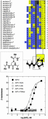
Sequence, hydrophobicity, and structure of various known and predicted AIPs. (A) At the top is displayed the sequences and hydrophobicity profiles of the 24 known or predicted AIPs. Most or all of the staphylococcal AIPs are 7-10 aa in length. In the alignment, hydrophilic (polar) residues are colored blue, and hydrophobic residues (nonpolar) are colored yellow. A shaded box marks the conserved cysteine, which is always 5 aa from the C terminus, and a black outline traces the conserved hydrophobic macrocycle residues. N3 in AIP-II and D5 and Y5 in AIP-I and -IV are highlighted because they are critical for activation (13, 20). Note that AIPs I and IV differ by only this amino acid and that Staphylococcus intermedius contains a serine in place of the conserved cysteine. Sequences marked with an asterisk are those that have been confirmed by in vitro synthesis and mass spectroscopy (11, 13-15, 36) (M. Kalkum, personal communication). The rest are predicted from the corresponding agrD sequences (12). Because of variation in the N-terminal tail region, most of the unconfirmed AIPs have been given an arbitrary length of 9 aa. Below this is a schematic model of a generic 9-aa AIP and a magnified view of the thiolactone bond. As a point of reference, highlighted residues are identified on the AIP model. (B) A representative dose-response plot with different AIP-II analogs against the AgrC-IV::III chimera demonstrates the importance of the conserved hydrophobic macrocycle residues in receptor recognition. The x axis represents the concentration of AIP, and the y axis indicates raw β-lactamase activity.
Fig. 4.
Fig. 4.
Hypothetical cartoon model illustrating activation and inhibition of AgrC. Binding of diverse but structurally similar AIPs to AgrC is driven by the hydrophobic patch (two circles) in the AIP endocycle region with an undisclosed hydrophobic binding pocket (shaded in yellow). The sharp features on the AIP represent specific molecular determinants for activation [N3 in AIP-II and D/Y5 in AIP-I and -IV (13, 20)] recognized by he distal subdomain of the receptor (shaded in blue). Activation is accompanied by molecular rearrangements in the receptor and/or AIP that propagate the signal to activate the HK domain and the subsequent phosphorelay to AgrA. Occupancy of a noncognate or inhibitory AIP lacking the requirements for activation results in competitive inhibition.

References

    1. Wolanin, P. M., Tomason, P. A. & Stock, J. B. (2002) Genome Biol. 3, 3013.1-3013.8. - PMC - PubMed
    1. Novick, R. P. & Muir, T. W. (1999) Curr. Opin. Microbiol. 2, 40-45. - PubMed
    1. Kleerebezem, M., Quadri, L. E., Kuipers, O. P. & de Vos, W. M. (1997) Mol. Microbiol. 24, 895-904. - PubMed
    1. Ji, G., Beavis, R. C. & Novick, R. P. (1995) Proc. Natl. Acad. Sci. USA 92, 12055-12059. - PMC - PubMed
    1. Nakayama, J., Cao, Y., Horii, T., Sakuda, S., Akkermans, A. D., de Vos, W. M. & Nagasawa, H. (2001) Mol. Microbiol. 41, 145-154. - PubMed

Publication types

MeSH terms

LinkOut - more resources