. 2004 Nov 15;173(10):6134-42.
doi: 10.4049/jimmunol.173.10.6134.

Human ribonuclease A superfamily members, eosinophil-derived neurotoxin and pancreatic ribonuclease, induce dendritic cell maturation and activation

Affiliations

Human ribonuclease A superfamily members, eosinophil-derived neurotoxin and pancreatic ribonuclease, induce dendritic cell maturation and activation

De Yang et al. J Immunol. .

Abstract

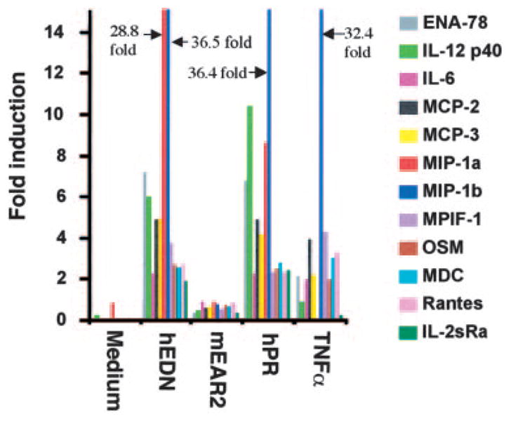
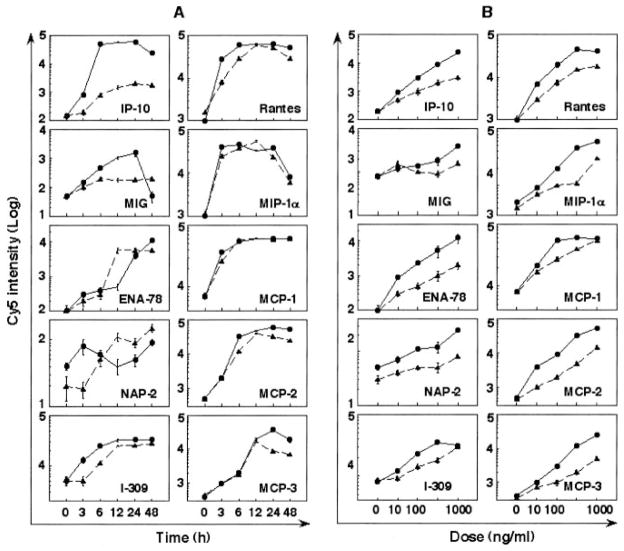
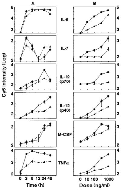
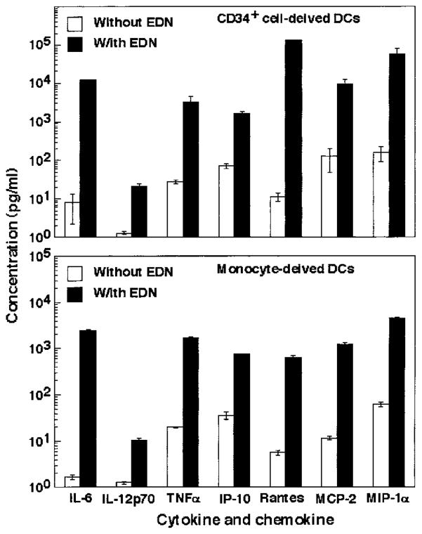
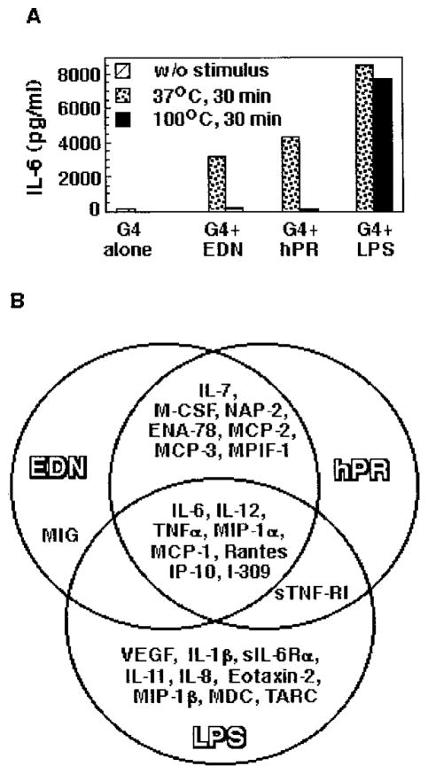
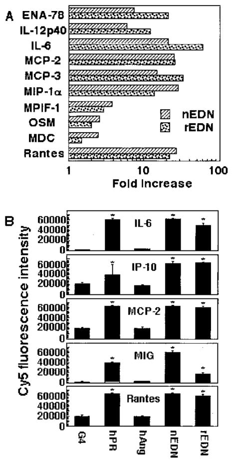
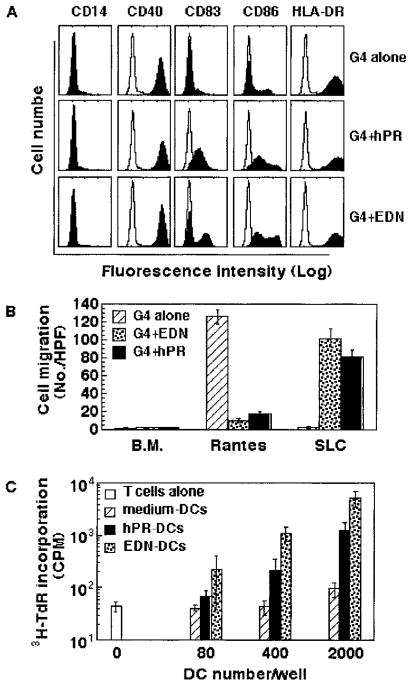
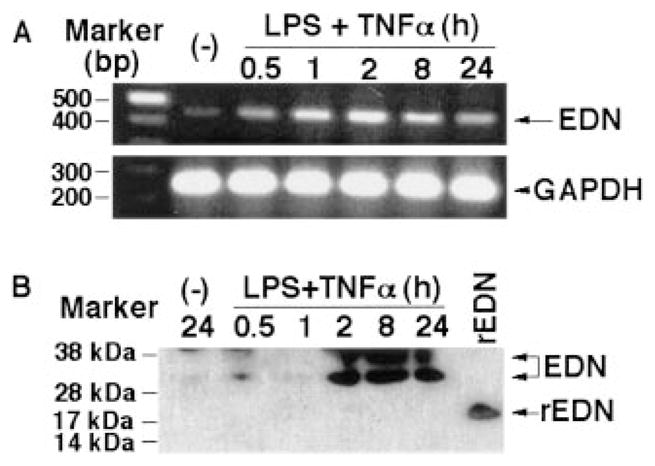
A number of mammalian antimicrobial proteins produced by neutrophils and cells of epithelial origin have chemotactic and activating effects on host cells, including cells of the immune system. Eosinophil granules contain an antimicrobial protein known as eosinophil-derived neurotoxin (EDN), which belongs to the RNase A superfamily. EDN has antiviral and chemotactic activities in vitro. In this study, we show that EDN, and to a lesser extent human pancreatic RNase (hPR), another RNase A superfamily member, activates human dendritic cells (DCs), leading to the production of a variety of inflammatory cytokines, chemokines, growth factors, and soluble receptors. Human angiogenin, a RNase evolutionarily more distant to EDN and hPR, did not display such activating effects. Additionally, EDN and hPR also induced phenotypic and functional maturation DCs. These RNases were as efficacious as TNF-alpha, but induced a different set of cytokine mediators. Furthermore, EDN production by human macrophages could be induced by proinflammatory stimuli. The results reveal the DC-activating activity of EDN and hPR and suggest that they are likely participants of inflammatory and immune responses. A number of endogenous mediators in addition to EDN have been reported to have both chemotactic and activating effects on APCs, and can thus amplify innate and Ag-specific immune responses to danger signals. We therefore propose these mediators be considered as endogenous multifunctional immune alarmins.


Figures

FIGURE 1
FIGURE 1
EDN and hPR induction of soluble mediator production by DCs. CD34+ progenitor-derived iDCs (106/ml) were cultured in G4 medium alone or in the presence of EDN (1 μg/ml), mEAR2 (1 μg/ml), hPR (1 μg/ml), or TNF-α (100 ng/ml) for 16 h at 37°C in humidified air containing 5% CO2. A dozen of soluble mediators in the culture supernatants was measured by RCA immunoassay, as described in Materials and Methods. The results are shown as fold induction, which, for a given soluble mediator, was calculated by the formula: amount produced by DCs in the presence of a stimulant/amount produced by DCs in G4 medium alone. IL-2sRa: soluble IL-2R α-chain.
FIGURE 2
FIGURE 2
Chemokine production induced by EDN (●) and hPR (▲) from CD34+ progenitor-derived DCs. A, Time course; iDCs (106/ml) were cultured in G4 medium containing 1 μ g/ml EDN or hPR for various time points, as specified. B, Dose response; iDCs (106/ml) were cultured for 48 h in G4 medium in the absence or presence of various concentrations of EDN or hPR, as specified. The culture supernatants were collected and measured by RCA immunoassay, as described in Materials and Methods.
FIGURE 3
FIGURE 3
Cytokine induction by EDN (●) and hPR (▲) from CD34+ progenitor-derived DCs. iDCs (106/ml) were cultured in G4 medium containing 1 μg/ml recombinant EDN or hPR for various times (A, time course) or for 48 h in G4 medium containing various concentrations of EDN or hPR (B, dose response). The culture supernatants were collected and measured by RCA immunoassay. Five cytokines induced by EDN or hPR are shown.
FIGURE 4
FIGURE 4
ELISA confirmation of EDN-stimulated DC production of cytokines and chemokines. iDCs generated from either bone marrow CD34+ progenitors (upper panel) or monocytes (lower panel) were cultured in triplicate in the absence (□) or presence (■) of EDN at 1 μg/ml for 48 h. The supernatants were harvested for performance of ELISA measurement on seven mediators, as indicated. Shown is the average (mean ± SD) of mediator production by CD34+ cell-derived DCs from two (upper panel) and monocyte-derived DC from four (lower panel) individuals.
FIGURE 5
FIGURE 5
A, The effect of heating on the IL-6-inducing capacity of recombinant EDN or hPR. EDN, hPR, and LPS samples were placed at 37°C or heated at 100°C for 30 min, followed by rapid cooling down on ice before addition to CD34+ progenitor-derived iDCs (106/ml). The final concentration for EDN, hPR, and LPS was 1 μg/ml, 1 μg/ml, and 100 ng/ml, respectively. After incubation at 37°C for 24 h in humidified air containing 5% CO2, the supernatants were collected and measured by ELISA for IL-6 production. B, Comparison of production of mediators by CD34+ progenitor-derived iDCs cultured for 48 h in the presence of recombinant EDN (1 μg/ml), hPR (1 μg/ml), and LPS (100 ng/ml). Only those mediators increased by ≥3-fold over control levels were included.
FIGURE 6
FIGURE 6
Comparison of the production of selected cytokines and chemokines by DCs in response to various RNase samples. A, CD34+ progenitor-derived iDCs were incubated in G4 medium alone or in the presence of natural (nEDN) or rEDN at 1 μg/ml at 37°C in humidified air containing 5% CO2. After 48 h of culture, the supernatants were assayed for 10 selected cytokines and chemokines by RCA immunoassay. The induction of cytokines and chemokines is presented as fold increase, which was calculated by the following formula: fold increase = Cy5 fluorescence in the presence of EDN or human pancreatic RNase/Cy5 fluorescence in the absence of EDN or hPR. B, Monocyte-derived iDCs were incubated in triplicates in G4 medium alone or in the presence of hPR, hANG, and natural and recombinant EDNs (at 1 μg/ml) at 37°C for 48 h in humidified air containing 5% CO2. The production of IL-6 and IL-4 chemokines in the culture supernatants was measured by RCA immunoassay and shown as the average (mean ± SEM) Cy5 fluorescence intensity. *, p < 0.05 when compared with G4-treated group.
FIGURE 7
FIGURE 7
Induction of DC maturation by hPR and EDN. Monocyte-derived iDCs were incubated in G4 medium alone or in the presence of recombinant hPR or EDNs (at 1 μg/ml) at 37°C for 48 in humidified air containing 5% CO2. The cells were collected and analyzed by flow cytometry, chemotaxis assay, or allogeneic MLR, as described in Materials and Methods. A, Surface expression of CD14, CD40, CD83, CD86, and HLA-DR by DCs cultured under conditions, as specified. B, Comparison of the chemotatic responses of DCs cultured in the absence or presence of 1 μg/ml EDN or hPR at 37°C for 48 h to optimal concentration (100 ng/ml) of RANTES or SLC. C, Stimulation of the proliferation of allogeneic human peripheral blood T lymphocytes by EDN- or hPR-treated DCs. T cells (105/well) were cultured in triplicate alone or with treated DCs at concentrations specified in 96-well plates for 7 days with the addition of [3H]TdR (0.5 μCi/well) in the last 8 h of incubation. The cells were harvested and measured for the incorporation of [3H]TdR (mean ± SD).
FIGURE 8
FIGURE 8
Up-regulation of EDN expression by macrophages in response to TNF-α and LPS. A, Human monocyte-derived macrophages were incubated in RPMI 1640 medium alone or in the presence of a combination of LPS and human rTNF-α (each at 100 ng/ml) at 37°C in humidified air containing 5% CO2 for a period of time, as indicated. Subsequently, total RNA was extracted and used as the template for the amplification of EDN or GAPDH by RT-PCR, as described in Materials and Methods. Upper and lower panels, EDN and GAPDH, respectively. B, Macrophage culture supernatant was harvested under conditions as specified, and the amount equivalent to 20 μg of protein was loaded onto each lane of a SDS-PAGE gel. rEDN (100 ng) generated in E. coli was used as a control (last lane). After transfer, the polyvinylidene difluoride membrane was blotted with rabbit anti-EDN polyclonal Ab, as detailed in Materials and Methods.

References

    1. Zasloff M. Antimicrobial peptides of multicellular organisms. Nature. 2002;415:389. - PubMed
    1. Yang D, Chertov O, Oppenheim JJ. The role of mammalian antimicrobial peptides and proteins in awakening of innate host defenses and adaptive immunity. Cell Mol Life Sci. 2001;58:978. - PMC - PubMed
    1. Scott MG, Hancock REW. Cationic antimicrobial peptides and their multifunctional role in the immune system. Crit Rev Immunol. 2002;20:407. - PubMed
    1. Chertov O, Michiel DF, Xu L, Wang JM, Tani K, Murphy WJ, Longo DL, Taub DD, Oppenheim JJ. Identification of defensin-1, defensin-2, and CAP37/azurocidin as T-cell chemoattractant proteins released from interleukin-8-stimulated neutrophils. J Biol Chem. 1996;271:2935. - PubMed
    1. Yang D, Chen Q, Chertov O, Oppenheim JJ. Human neutrophil defensins selectively chemoattract naive T and immature dendritic cells. J Leukocyte Biol. 2000;68:9. - PubMed

Publication types

MeSH terms