

Review
doi: 10.1126/science.1099993.
Autophagy in health and disease: a double-edged sword
Affiliations
- PMID: 15528435
- PMCID: PMC1705980
- DOI: 10.1126/science.1099993
Item in Clipboard
Review
Autophagy in health and disease: a double-edged sword
Science.
.
Abstract
Autophagy, the process by which cells recycle cytoplasm and dispose of excess or defective organelles, has entered the research spotlight largely owing to the discovery of the protein components that drive this process. Identifying the autophagy genes in yeast and finding orthologs in other organisms reveals the conservation of the mechanism of autophagy in eukaryotes and allows the use of molecular genetics and biology in different model systems to study this process. By mostly morphological studies, autophagy has been linked to disease processes. Whether autophagy protects from or causes disease is unclear. Here, we summarize current knowledge about the role of autophagy in disease and health.
Figures
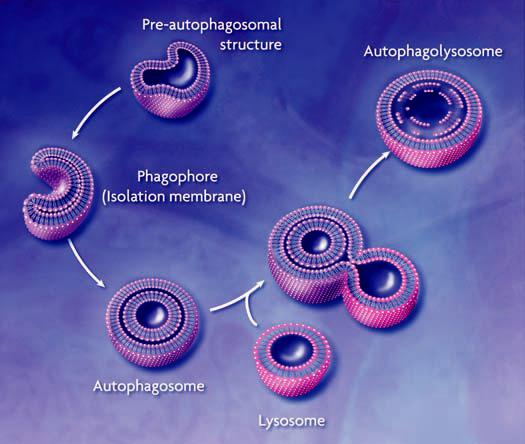
Conceptual model of macroautophagy. A sequestering membrane, termed a phagophore or isolation membrane, forms from the pre-autophagosomal structure. The source of the membrane is unknown but probably includes the endoplasmic reticulum and early secretory pathway. The isolation membrane enwraps cytosol and organelles; on completion, a double-membrane vesicle, the autophagosome or autophagic vacuole, is formed. The autophagosome acquires hydrolytic enzymes by fusing with the lysosome to generate an autophagolysosome, and the inner vesicle of the autophagosome is released into the lumen. The resulting autophagic body is broken down, allowing access to, and degradation and recycling of, the cargo.
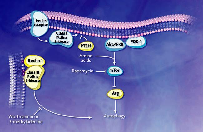
Schematic model of autophagic regulation. Stimulation of the class I PtdIns 3-kinase at the plasma membrane through the insulin receptor results in the generation of PtdIns(3,4)P2 and PtdIns(3,4,5)P3 (dark pink circles). These phosphoinositides allow binding and activation of Akt/PKB and its activator PDK-1. Along with amino acids, Akt/PKB activates mTor (additional components in this pathway are not depicted). Subsequent phosphorylation of a downstream effector, possibly analogous to Atg1 or other ATG gene products as demonstrated in yeast, inhibits autophagy. PTEN dephosphorylates 3′ phosphoinositides and antagonizes the action of class I PtdIns 3-kinase. A class III PtdIns 3-kinase complex, which includes Beclin 1/Atg6, generates PtdIns(3)P (purple circles) to control the membrane dynamics that are associated with autophagosome formation. Rapamycin inhibits mTor, while wortmannin and 3-methyladenine inhibit the class III PtdIns 3-kinase; the effect is to induce or inhibit autophagy, respectively. Autophagy is also regulated through heterotrimeric G proteins and other kinases and phosphatases that are not depicted.
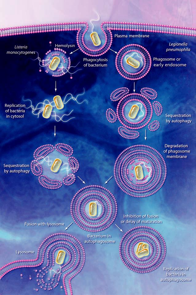
Action and subversion of autophagy during bacterial infection. Bacteria may be taken up by phagocytosis and the resulting phagosome can fuse with endosomes and then the lysosome; the bacteria are then degraded within the phagolysosome (not shown). Some pathogens such as L. monocytogenes, escape this pathway by lysing the phagosome membrane. The bacteria may subsequently become targets for autophagy. In the case of L. pneumophila, P. gingivalis, and B. abortus, the phagosome either fuses with, or becomes sequestered within, the autophagosome. Inhibition of autophagosome maturation or a delay in fusion with the lysosome, dependent on a type IV secretion system, allows the bacteria to replicate within the autophagosome and/or autophagolysosome (in the case of L. pneumophila) and possibly become resistant to lysosomal degradation. In addition, degradation of host cell proteins within the late autophagosome or autophagolysosome may supply the nutrients needed for growth of the pathogen.
References
Publication types
MeSH terms
Grants and funding
LinkOut - more resources
Full Text Sources
Other Literature Sources
Molecular Biology Databases
