. 2004 Nov;70(11):6826-33.
doi: 10.1128/AEM.70.11.6826-6833.2004.

Intracellular proliferation of Legionella pneumophila in Hartmannella vermiformis in aquatic biofilms grown on plasticized polyvinyl chloride

Affiliations

Intracellular proliferation of Legionella pneumophila in Hartmannella vermiformis in aquatic biofilms grown on plasticized polyvinyl chloride

Melanie W Kuiper et al. Appl Environ Microbiol. 2004 Nov.

Abstract

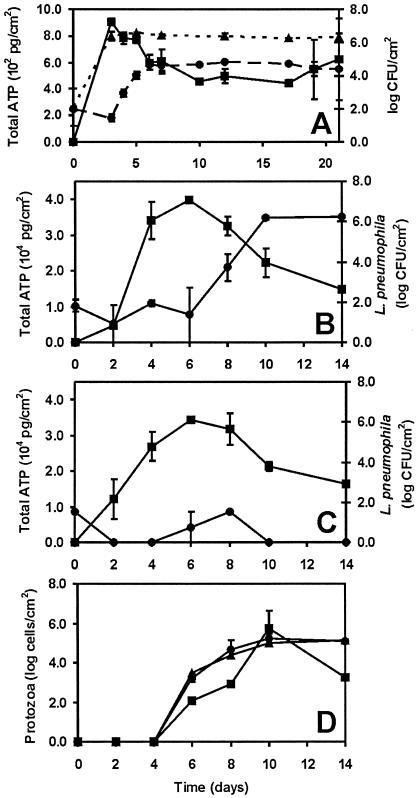
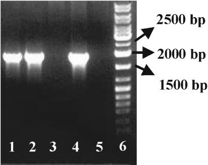
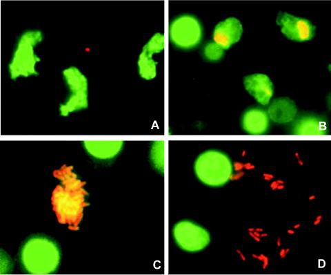
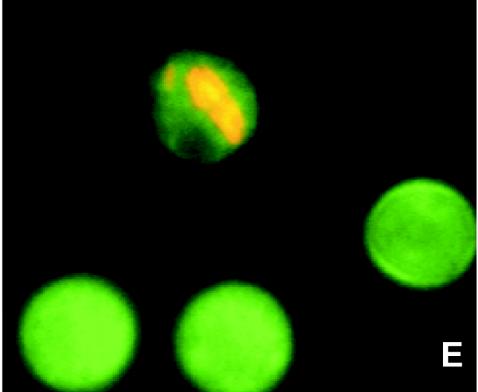
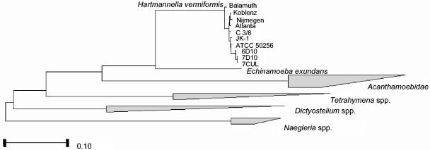
The need for protozoa for the proliferation of Legionella pneumophila in aquatic habitats is still not fully understood and is even questioned by some investigators. This study shows the in vivo growth of L. pneumophila in protozoa in aquatic biofilms developing at high concentrations on plasticized polyvinyl chloride in a batch system with autoclaved tap water. The inoculum, a mixed microbial community including indigenous L. pneumophila originating from a tap water system, was added in an unfiltered as well as filtered (cellulose nitrate, 3.0-microm pore size) state. Both the attached and suspended biomasses were examined for their total amounts of ATP, for culturable L. pneumophila, and for their concentrations of protozoa. L. pneumophila grew to high numbers (6.3 log CFU/cm2) only in flasks with an unfiltered inoculum. Filtration obviously removed the growth-supporting factor, but it did not affect biofilm formation, as determined by measuring ATP. Cultivation, direct counting, and 18S ribosomal DNA-targeted PCR with subsequent sequencing revealed the presence of Hartmannella vermiformis in all flasks in which L. pneumophila multiplied and also when cycloheximide had been added. Fluorescent in situ hybridization clearly demonstrated the intracellular growth of L. pneumophila in trophozoites of H. vermiformis, with 25.9% +/- 10.5% of the trophozoites containing L. pneumophila on day 10 and >90% containing L. pneumophila on day 14. Calculations confirmed that intracellular growth was most likely the only way for L. pneumophila to proliferate within the biofilm. Higher biofilm concentrations, measured as amounts of ATP, gave higher L. pneumophila concentrations, and therefore the growth of L. pneumophila within engineered water systems can be limited by controlling biofilm formation.


Figures

FIG. 1.
FIG. 1.
(A) Concentrations of total ATP (▪), L. pneumophila (•), and heterotrophic bacteria (▴) in flasks with the unfiltered inoculum and PVCu as the biofilm carrier. Each point represents the mean concentration from two experiments, and bars indicate standard errors. (B) Concentrations of total ATP (▪) and L. pneumophila (•) in flasks with the unfiltered inoculum and PVCp as the biofilm carrier. Each point represents the mean concentration from two experiments, and bars indicate standard errors. (C) Concentrations of total ATP (▪) and L. pneumophila (•) in flasks with the filtered inoculum and PVCp as the biofilm carrier. Each point represents the mean concentration from two experiments, and bars indicate standard errors. (D) Protozoon concentrations in flasks with the unfiltered inoculum. Squares (▪) represent the concentrations determined by the cultivation method; circles (•) and triangles (▴) represent the concentrations determined by direct counting methods using primulin and FISH, respectively. Bars indicate standard errors. No protozoa were detected in flasks with the filtered inoculum.
FIG. 2.
FIG. 2.
Detection of eukaryotic DNA. Lane 1, DNA from biofilm from flasks with unfiltered inoculum in the presence of 100 μM cycloheximide; lanes 2 and 4, DNAs from biofilms from flasks with unfiltered inoculum; lanes 3 and 5, DNAs from biofilms from flasks with filtered inoculum; lane 6, marker (sm 0333 gene ruler DNA ladder mix; Fermentas, St. Leon-Rot, Germany).
FIG. 3.
FIG. 3.
Different stages of intracellular proliferation of L. pneumophila within amoebae. All images were made by using material from the biofilm phase, since amoebae were mainly present in this phase. (A) Trophozoite amoebae on day 6 of the experiment. Magnification, ×1,000. (B) Trophozoite amoebae that were just infected with L. pneumophila (day 10). Magnification, ×1,000. (C) Heavily infected amoeba (day 14). Magnification, ×1,600. (D) Free L. pneumophila bacteria, just escaped from a lysed amoeba (day 14). Magnification, ×1,600. (E) Cysts of amoebae, with one infected by L. pneumophila (day 14). Magnification, ×1,600.
FIG. 3.
FIG. 3.
Different stages of intracellular proliferation of L. pneumophila within amoebae. All images were made by using material from the biofilm phase, since amoebae were mainly present in this phase. (A) Trophozoite amoebae on day 6 of the experiment. Magnification, ×1,000. (B) Trophozoite amoebae that were just infected with L. pneumophila (day 10). Magnification, ×1,000. (C) Heavily infected amoeba (day 14). Magnification, ×1,600. (D) Free L. pneumophila bacteria, just escaped from a lysed amoeba (day 14). Magnification, ×1,600. (E) Cysts of amoebae, with one infected by L. pneumophila (day 14). Magnification, ×1,600.
FIG. 4.
FIG. 4.
Phylogenetic tree from 18S rRNA gene sequences showing the relationships of the examined sequences to those of other protozoa in which intracellular growth of L. pneumophila was shown (12, 15, 21, 39). Different trees were calculated, and all trees gave the same consensus, as shown in the figure. Bar, 10% sequence divergence. Strains Koblenz (X75514), Nijmegen (X75515), and Atlanta (X75513) were previously sequenced by Weekers et al. (49), strain Balamuth (ATCC 30966, M95168) was sequenced by Gunderson et al. (20), strain C3/8 (AF426157) was sequenced by Walochnik et al. (48), and strains JK-1 and ATCC 50256 were sequenced by Kuchta et al. (24). Clones: 6D10, KWR1; 7D10, KWR2; 7CUL, KWR3.

References

    1. Amann, R. I., B. J. Binder, R. J. Olson, S. W. Chisholm, D. Richard, and D. A. Stahl. 1990. Combination of 16S rRNA-targeted oligonucleotide probes with flow cytometry for analyzing mixed microbial populations. Appl. Environ. Microbiol. 56:1919-1925. - PMC - PubMed
    1. Amaral Zettler, L. A., T. A. Nerad, C. J. O'Kelly, M. T. Peglar, P. M. Gillevet, J. D. Silberman, and M. L. Sogin. 2000. A molecular reassessment of the leptomyxid amoebae. Protist 151:275-282. - PubMed
    1. Arndt, H. 1993. A critical review of the importance of rhizopods (naked and testate amoebae) and actinopods (heliozoa) in lake plankton. Mar. Microb. Food Webs 7:3-29.
    1. Baldock, B. M., J. H. Baker, and M. A. Sleigh. 1980. Laboratory growth rates of six species of freshwater gymnamoebia. Oecologia 47:156-159. - PubMed
    1. Barbaree, J. M., B. S. Fields, J. C. Feeley, G. W. Gorman, and W. T. Martin. 1985. Isolation of protozoa from water associated with a legionellosis outbreak and demonstration of intracellular multiplication of Legionella pneumophila. Appl. Environ. Microbiol. 51:422-424. - PMC - PubMed

Publication types

Associated data

LinkOut - more resources