. 2004 Nov;50(11):3541-8.
doi: 10.1002/art.20601.

Role of NF-kappaB transcription factors in antiinflammatory and proinflammatory actions of mechanical signals

Affiliations

Role of NF-kappaB transcription factors in antiinflammatory and proinflammatory actions of mechanical signals

Sudha Agarwal et al. Arthritis Rheum. 2004 Nov.

Abstract

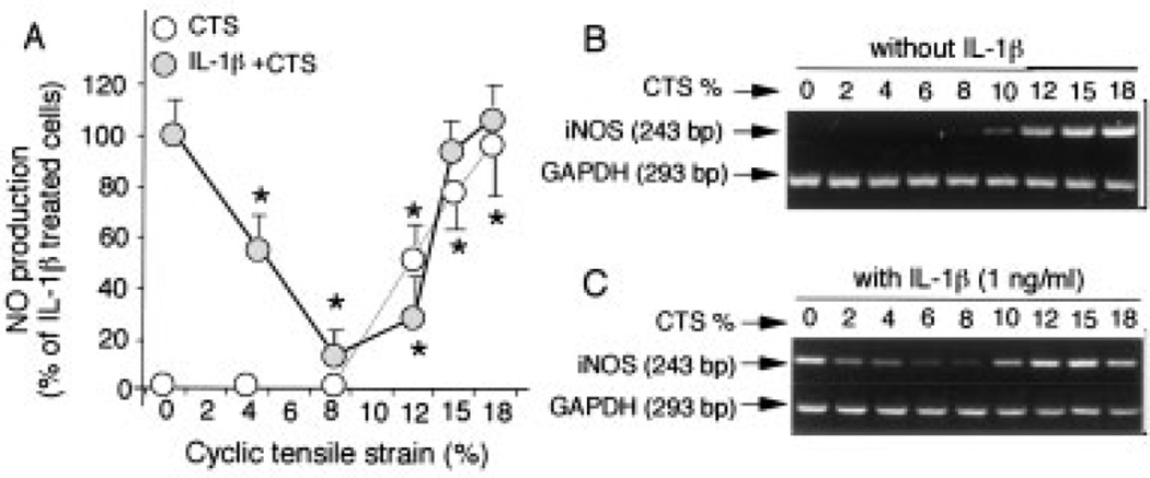
Objective: The mechanisms by which chondrocytes convert biomechanical signals into intracellular biochemical events are not well understood. In this study, we sought to determine the intracellular mechanisms of the magnitude-dependent actions of mechanical signals.

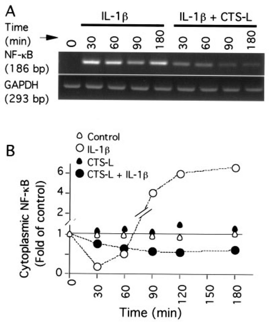
Methods: Chondrocytes isolated from rabbit articular cartilage were grown on flexible membranes. Cells were subjected to cyclic tensile strain (CTS) of various magnitudes in the presence or absence of interleukin-1beta (IL-1beta), which was used as a proinflammatory signal for designated time intervals. The regulation of NF-kappaB was measured by reverse transcriptase-polymerase chain reaction, electrophoretic mobility shift assay, and immunofluorescence.

Results: CTS of low magnitudes (4-8% equibiaxial strain) was a potent inhibitor of IL-1beta-dependent NF-kappaB nuclear translocation. Cytoplasmic retention of NF-kappaB and reduction of its synthesis led to sustained suppression of proinflammatory gene induction. In contrast, proinflammatory signals generated by CTS of high magnitudes (15-18% equibiaxial strain) mimicked the actions of IL-1beta and induced rapid nuclear translocation of NF-kappaB subunits p65 and p50.

Conclusion: Magnitude-dependent signals of mechanical strain utilize the NF-kappaB transcription factors as common elements to abrogate or aggravate proinflammatory responses. Furthermore, the intracellular events induced by mechanical overload are similar to those that are initiated by proinflammatory cytokines in arthritis.


Figures

Figure 1
Figure 1
Magnitude-dependent response of chondrocytes to mechanical signals. A, Regulation of nitric oxide (NO) production by various magnitudes of cyclic tensile strain (CTS) in the presence and absence of interleukin-1β (IL-1β) (1.0 ng/ml). Accumulation of NO in the culture supernatants was assessed after 24 hours. Values are the mean and SEM of triplicate determinations. * = P ≤ 0.05 between unstretched cells and cells subjected to CTS in the absence and presence of IL-1β, by Student’s t-test. B and C, The regulation of inducible NO synthase (iNOS) mRNA expression by various magnitudes of CTS was determined in B, the absence and C, the presence of IL-1β (1.0 ng/ml). Expression of iNOS mRNA was measured by reverse transcriptase–polymerase chain reaction after 4 hours. Representative results from 1 of 3 separate experiments are shown.
Figure 2
Figure 2
Abrogation of interleukin-1β (IL-1β)–induced nuclear translocation of NF-κB by cyclic tensile strain of low magnitudes (CTS-L). A, Nuclear proteins of untreated cells (control) and cells treated for 15, 30, 60, or 90 minutes with IL-1β (1.0 ng/ml) and/or CTS-L (6%) were extracted. Subsequently, the presence of NF-κB in the nuclear extract was determined by electrophoretic mobility shift assay (EMSA). B, Quantitative analysis of net 32P associated with each band from the EMSA gel shown in A was performed by scintillation counting to assess NF-κB nuclear translocation. C, NF-κB subunits involved in the actions of CTS-L were analyzed by supershift EMSA (SS-EMSA) using antibodies against p65 (RelA) and p50 subunits of NF-κB. D, Chondrocytes were treated for 30, 60, 120, or 180 minutes with IL-1β (1.0 ng/ml) and/or CTS-L (6%). Untreated cells were used as controls. Nuclear translocation of NF-κB was assessed by immunofluorescence staining. NF-κB was stained with rabbit anti–NF-κB p65 IgG, with Cy3-conjugated goat anti-rabbit IgG as secondary antibody (red). Cellular β-actin was stained with fluorescein isothiocyanate–conjugated phalloidin (green). Representative results from 1 of 3 separate experiments are shown.
Figure 3
Figure 3
Inhibition of IL-1β–induced NF-κB mRNA expression and NF-κB synthesis by CTS-L. A, IL-1β–induced NF-κB p65 mRNA expression over a period of 30–180 minutes in the absence and presence of CTS-L (6%) was analyzed by reverse transcriptase–polymerase chain reaction. B, Cytoplasmic NF-κB p65 in untreated cells and cells treated with IL-1β (1.0 ng/ml) and/or CTS-L (6%) over a period of 30–180 minutes was determined by densitometric analysis of Western blots. Representative results from 1 of at least 3 separate experiments are shown. See Figure 2 for definitions.
Figure 4
Figure 4
Induction of NF-κB nuclear translocation and synthesis by CTS of high magnitudes (CTS-H). A, Nuclear proteins of untreated cells (control) and cells treated for 30, 60, 120, or 180 minutes with IL-1β (1.0 ng/ml) and/or CTS-H (15%) were extracted. Subsequently, the presence of NF-κB in the nuclear extract was determined by EMSA. Quantitative analysis of net 32P associated with each band determined by EMSA was performed to assess NF-κB nuclear translocation. Each point is the mean of triplicate values. B, NF-κB p65 mRNA expression induced by CTS-H (15%) and/or IL-1β (1.0 ng/ml) over a period of 180 minutes was analyzed by reverse transcriptase–polymerase chain reaction. C, NF-κB subunits involved in CTS-H actions (15%, 30 minutes) were analyzed by supershift EMSA using antibodies against p65, p50, p52, RelB, and c-Rel subunits of NF-κB. D, Immunofluorescence staining of chondrocytes subjected to CTS-H (15%) for 30, 60, or 120 minutes was performed in the presence or absence of IL-1β (1.0 ng/ml). Untreated cells were used as controls. NF-κB was stained with rabbit anti–NF-κB p65 IgG, with Cy3-conjugated goat anti-rabbit IgG as secondary antibody (red). Cellular β-actin was stained by fluorescein isothiocyanate–conjugated phalloidin (green). Representative results from 1 of 3 separate experiments are shown. See Figure 2 for other definitions.
Figure 5
Figure 5
Abrogation of CTS of high magnitudes (CTS-H)–induced nuclear translocation of NF-κB by caffeic acid phenethyl ester (CAPE). A, Induction of CTS-H–dependent nuclear translocation of NF-κB was inhibited by CAPE over a period of 30–180 minutes. Chondrocytes were either untreated or were treated with CAPE (100 µM) for 10 minutes prior to being subjected to CTS-H (15%) in the presence or absence of IL-1β (1.0 ng/ml). Subsequently, the presence of NF-κB in the nuclear extract was determined by EMSA. The radioactivity associated with bands determined by EMSA was measured with a scintillation counter. Values are the mean and SEM of triplicate determinations. B, Effect of various concentrations of CAPE on inducible nitric oxide synthase (iNOS) mRNA expression induced by IL-1β and/or CTS-H. Chondrocytes were exposed to various concentrations of CAPE (0, 5, 25, 50, or 100 µM) for 10 minutes prior to being treated with IL-1β (1.0 ng/ml) and/or CTS-H (15%) for 4 hours. The iNOS mRNA expression was analyzed by reverse transcriptase–polymerase chain reaction (RT-PCR). C, Densitometric analysis of the RT-PCR gels shown in B. Representative results from 1 of 3 separate experiments are shown. See Figure 2 for other definitions.

References

    1. Quinn TM, Allen RG, Schalet BJ, Perumbuli P, Hunziker EB. Matrix and cell injury due to sub-impact loading of adult bovine articular cartilage explants: effects of strain rate and peak stress. J Orthop Res. 2001;19:242–249. - PubMed
    1. Patwari P, Cook MN, DiMicco MA, Blake SM, James IE, Kumar S, et al. Proteoglycan degradation after injurious compression of bovine and human articular cartilage in vitro: interaction with exogenous cytokines. Arthritis Rheum. 2003;48:1292–1301. - PubMed
    1. Soltz MA, Ateshian GA. A Conewise Linear Elasticity mixture model for the analysis of tension-compression nonlinearity in articular cartilage. J Biomech Eng. 2000;122:576–586. - PMC - PubMed
    1. Huang CY, Soltz MA, Kopacz M, Mow VC, Ateshian GA. Experimental verification of the roles of intrinsic matrix viscoelasticity and tension-compression nonlinearity in the biphasic response of cartilage. J Biomech Eng. 2003;125:84–93. - PubMed
    1. Buschmann MD, Kim YJ, Wong M, Frank E, Hunziker EB, Grodzinsky AJ. Stimulation of aggrecan synthesis in cartilage explants by cyclic loading is localized to regions of high interstitial fluid flow. Arch Biochem Biophys. 1999;366:1–7. - PubMed

Publication types