

Radiation-force assisted targeting facilitates ultrasonic molecular imaging
- PMID: 15530249
- PMCID: PMC1356635
- DOI: 10.1162/15353500200404115
Radiation-force assisted targeting facilitates ultrasonic molecular imaging
Abstract
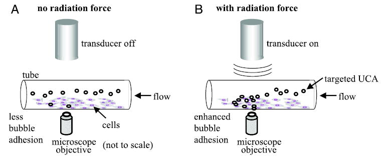
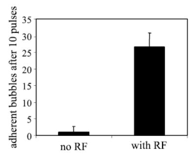
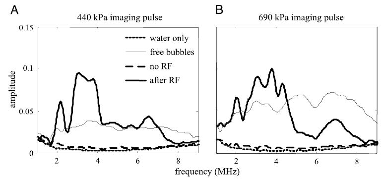
Ultrasonic molecular imaging employs contrast agents, such as microbubbles, nanoparticles, or liposomes, coated with ligands specific for receptors expressed on cells at sites of angiogenesis, inflammation, or thrombus. Concentration of these highly echogenic contrast agents at a target site enhances the ultrasound signal received from that site, promoting ultrasonic detection and analysis of disease states. In this article, we show that acoustic radiation force can be used to displace targeted contrast agents to a vessel wall, greatly increasing the number of agents binding to available surface receptors. We provide a theoretical evaluation of the magnitude of acoustic radiation force and show that it is possible to displace micron-sized agents physiologically relevant distances. Following this, we show in a series of experiments that acoustic radiation force can enhance the binding of targeted agents: The number of biotinylated microbubbles adherent to a synthetic vessel coated with avidin increases as much as 20-fold when acoustic radiation force is applied; the adhesion of contrast agents targeted to alpha(v)beta3 expressed on human umbilical vein endothelial cells increases 27-fold within a mimetic vessel when radiation force is applied; and finally, the image signal-to-noise ratio in a phantom vessel increases up to 25 dB using a combination of radiation force and a targeted contrast agent, over use of a targeted contrast agent alone.
Figures


Similar articles
-
A sensitive TLRH targeted imaging technique for ultrasonic molecular imaging.IEEE Trans Ultrason Ferroelectr Freq Control. 2010;57(2):305-16. doi: 10.1109/TUFFC.2010.1411. IEEE Trans Ultrason Ferroelectr Freq Control. 2010. PMID: 20178897 Free PMC article.
-
Unbinding of targeted ultrasound contrast agent microbubbles by secondary acoustic forces.Phys Med Biol. 2011 Oct 7;56(19):6161-77. doi: 10.1088/0031-9155/56/19/002. Epub 2011 Aug 30. Phys Med Biol. 2011. PMID: 21878709
-
Selective imaging of adherent targeted ultrasound contrast agents.Phys Med Biol. 2007 Apr 21;52(8):2055-72. doi: 10.1088/0031-9155/52/8/002. Epub 2007 Mar 20. Phys Med Biol. 2007. PMID: 17404455 Free PMC article.
-
[Ultrasound contrast agents--physical basics].Radiologe. 2005 Jun;45(6):503-12. doi: 10.1007/s00117-005-1188-z. Radiologe. 2005. PMID: 15809841 Review. German.
-
Bubble dynamics involved in ultrasonic imaging.Expert Rev Mol Diagn. 2006 May;6(3):493-502. doi: 10.1586/14737159.6.3.493. Expert Rev Mol Diagn. 2006. PMID: 16706749 Review.
Cited by
-
Ultrasound Molecular Imaging as a Potential Non-invasive Diagnosis to Detect the Margin of Hepatocarcinoma via CSF-1R Targeting.Front Bioeng Biotechnol. 2020 Jul 14;8:783. doi: 10.3389/fbioe.2020.00783. eCollection 2020. Front Bioeng Biotechnol. 2020. PMID: 32760707 Free PMC article.
-
Lung surfactant microbubbles increase lipophilic drug payload for ultrasound-targeted delivery.Theranostics. 2013 May 20;3(6):409-19. doi: 10.7150/thno.5616. Print 2013. Theranostics. 2013. PMID: 23781287 Free PMC article.
-
Influence of lipid shell physicochemical properties on ultrasound-induced microbubble destruction.IEEE Trans Ultrason Ferroelectr Freq Control. 2005 Nov;52(11):1992-2002. doi: 10.1109/tuffc.2005.1561668. IEEE Trans Ultrason Ferroelectr Freq Control. 2005. PMID: 16422411 Free PMC article.
-
Optical Verification of Microbubble Response to Acoustic Radiation Force in Large Vessels With In Vivo Results.Invest Radiol. 2015 Nov;50(11):772-84. doi: 10.1097/RLI.0000000000000185. Invest Radiol. 2015. PMID: 26135018 Free PMC article.
-
Combining microbubbles and ultrasound for drug delivery to brain tumors: current progress and overview.Theranostics. 2014 Feb 12;4(4):432-44. doi: 10.7150/thno.8074. eCollection 2014. Theranostics. 2014. PMID: 24578726 Free PMC article. Review.
References
-
- Lanza GM, Wickline SA. Targeted ultrasonic contrast agents for molecular imaging and therapy. Prog Cardiovasc Dis. 2001;44:13–31. - PubMed
-
- Lanza GM, Wallace KD, Scott MJ, Cacheris WP, Abendschein DR, Christy DH, Sharkey AM, Miller JG, Gaffney PJ, Wickline SA. A novel site-targeted ultrasonic contrast agent with broad biomedical application. Circulation. 1996;94:3334–3340. - PubMed
-
- Lindner JR. Evolving applications for contrast ultrasound. Am J Cardiol. 2002;90:72J–80J. - PubMed
-
- Dayton P, Klibanov A, Brandenburger G, Ferrara K. Acoustic radiation force in vivo: A mechanism to assist targeting of microbubbles. Ultrasound Med Biol. 1999;25:1195–1201. - PubMed
-
- Dayton PA, Ferrara KW. Targeted imaging using ultrasound. J Magn Reson Imaging. 2002;16:362–377. - PubMed
Publication types
MeSH terms
Substances
Grants and funding
LinkOut - more resources
Full Text Sources
Other Literature Sources
Miscellaneous
