

Review
doi: 10.1016/j.ceb.2004.09.012.
Misfolded proteins, endoplasmic reticulum stress and neurodegeneration
Affiliations
- PMID: 15530777
- PMCID: PMC3970707
- DOI: 10.1016/j.ceb.2004.09.012
Item in Clipboard
Review
Misfolded proteins, endoplasmic reticulum stress and neurodegeneration
Curr Opin Cell Biol.
2004 Dec.
Abstract
The accumulation of misfolded proteins (e.g. mutant or damaged proteins) triggers cellular stress responses that protect cells against the toxic buildup of such proteins. However, prolonged stress due to the buildup of these toxic proteins induces specific death pathways. Dissecting these pathways should be valuable in understanding the pathogenesis of, and ultimately in designing therapy for, neurodegenerative diseases that feature misfolded proteins.
Figures
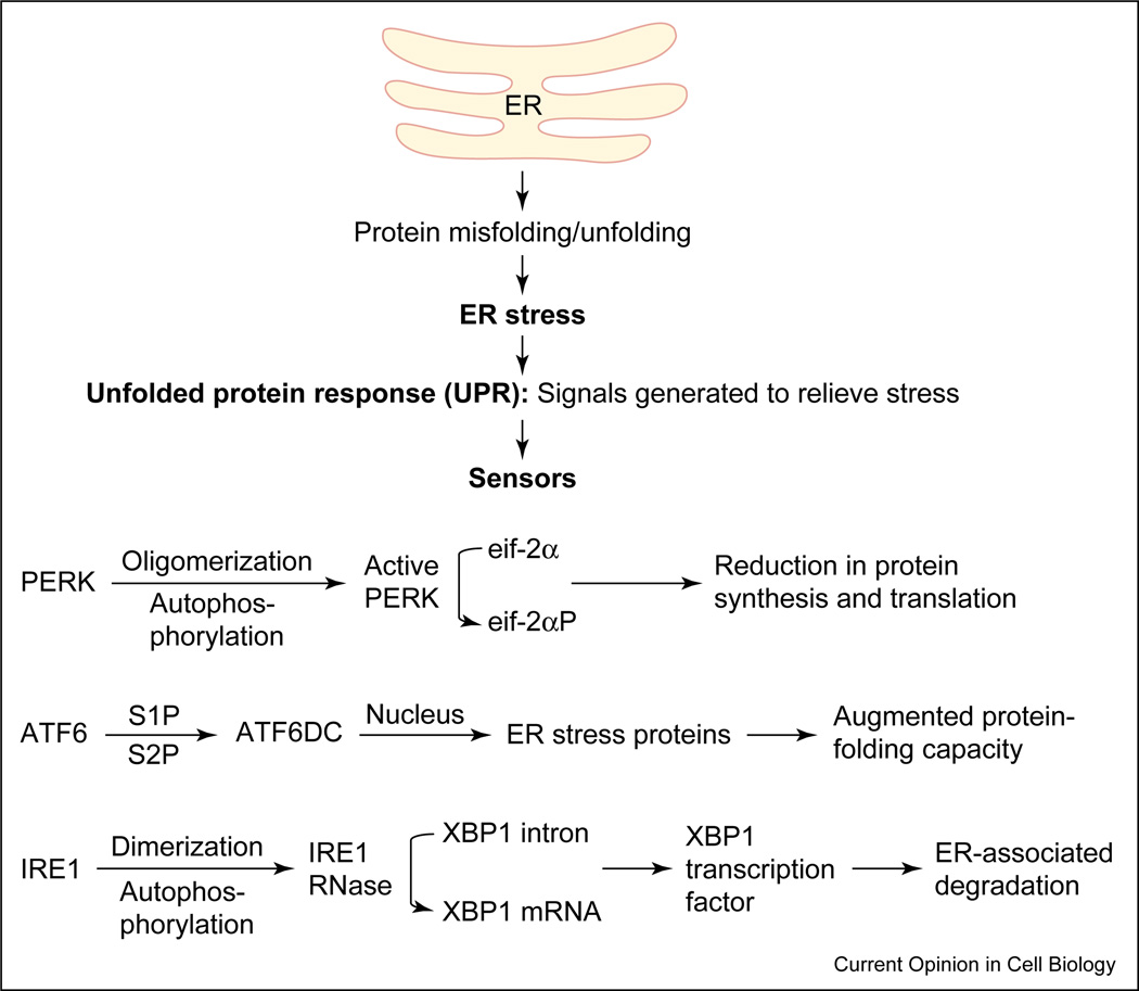
Accumulation of misfolded proteins in the ER can disrupt ER function resulting in ‘ER stress’. The ER responds by triggering specific signaling pathways including the UPR. The UPR is coordinately regulated by the three proximal sensors, IRE1, PERK and ATF6. The activation of all three proximal sensors results in reduction in the amount of new protein translocated into the ER lumen, increased degradation of ER-localized proteins and increased protein folding capacity of the ER. ATF6DC represents the 50kD cytosolic bZIP-containing fragment that translocates to the nucleus to activate transcription.

The UPR is negatively regulated by GRP78/Bip, which associates with the three proximal sensors, IRE1, PERK and ATF6. GRP78 binds to the luminal domains of IRE1 and PERK and prevents their dimerization and activation. GRP78 associates with ATF6 and prevents its translocation to the Golgi for further activation. In the presence of misfolded proteins, GRP78 dissociates from the sensors and binds the misfolded proteins, thus releasing the sensors from negative inhibition. The three sensors coordinately regulate the UPR through their various signaling pathways.
References
-
- Selkoe DJ. Folding proteins in fatal ways. Nature. 2003;426:900–904. - PubMed
-
- Taylor JP, Hardy J, Fischbeck KH. Toxic proteins in neurodegenerative disease. Science. 2002;296:1991–1995. - PubMed
-
- Kopito RR, Ron D. Conformational disease. Nat Cell Biol. 2000;2:E207–E209. - PubMed
-
- Dobson CM. Protein folding and misfolding. Nature. 2003;426:884–890. - PubMed
-
- Kopito RR. Aggresomes, inclusion bodies and protein aggregation. Trends Cell Biol. 2000;10:524–530. - PubMed
Publication types
MeSH terms
Substances
Grants and funding
LinkOut - more resources
Full Text Sources
Other Literature Sources
