. 2004 Dec 10;301(2):147-57.
doi: 10.1016/j.yexcr.2004.07.008.

Retinoids arrest breast cancer cell proliferation: retinoic acid selectively reduces the duration of receptor tyrosine kinase signaling

Affiliations

Retinoids arrest breast cancer cell proliferation: retinoic acid selectively reduces the duration of receptor tyrosine kinase signaling

Ann P Tighe et al. Exp Cell Res. .

Abstract

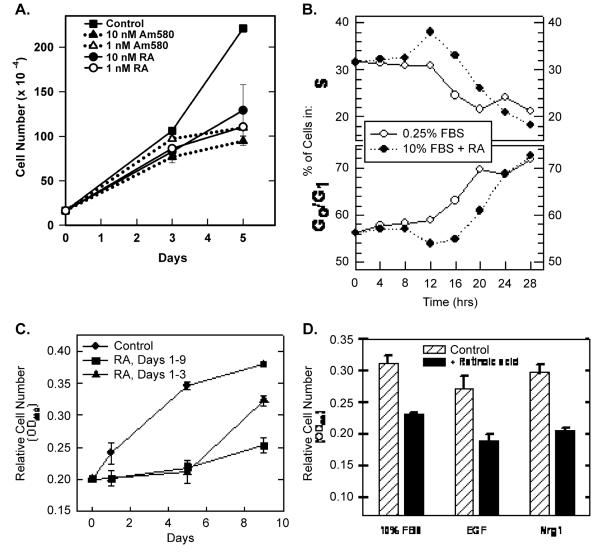
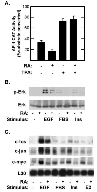
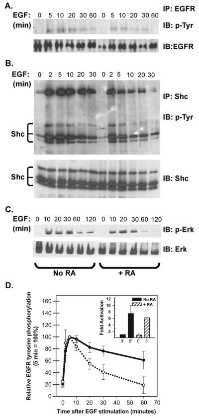
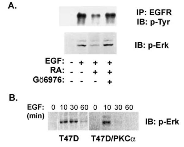
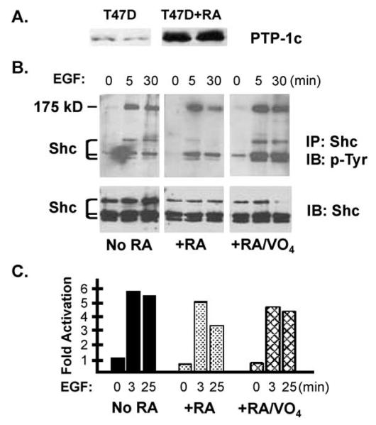
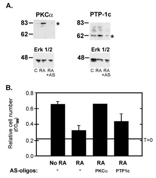
Retinoic acid (RA) induces cell cycle arrest of hormone-dependent human breast cancer (HBC) cells. Previously, we demonstrated that RA-induced growth arrest of T-47D HBC cells required the activity of the RA-induced protein kinase, protein kinase Calpha (PKCalpha) [J. Cell Physiol. 172 (1997) 306]. Here, we demonstrate that RA treatment of T-47D cells interfered with growth factor signaling to downstream, cytoplasmic and nuclear targets. RA treatment did not inhibit epidermal growth factor (EGF) receptor activation but resulted in rapid inactivation. The lack of sustained EGFR activation was associated with transient rather than sustained association of the EGFR with the Shc adaptor proteins and activation of Erk 1/2 and with compromised induction of expression of immediate early response genes. Inhibiting the activity of PKCalpha, a retinoic acid-induced target gene, prevented the effects of RA on cell proliferation and EGF signaling. Constitutive expression of PKCalpha, in the absence of RA, decreased cell proliferation and decreased EGF signaling. RA treatment increased steady-state levels of the protein tyrosine phosphatase PTP-1C and all measured effects of RA on EGF receptor function were reversed by the tyrosine phosphate inhibitor orthovanadate. These results indicate that RA-induced target genes, particularly PKCalpha, prevent sustained growth factor signaling, uncoupling activated receptor tyrosine kinases and nuclear targets that are required for cell cycle progression.


Figures

Fig. 1
Fig. 1
Retinoids reversibly inhibit T-47D cell proliferation. (A) T-47D cells (initial plating density = 1.5 × 10⁵ cells per 60-mm dish) were cultured in the presence of the indicated concentration of RA or the RARα-selective retinoid, Am580. Day 0 represents the time of retinoid addition. Cells were trypsinized and cell numbers determined 3 and 5 days later (data plotted ± SEM). Viability, as determined by trypan blue exclusion, was >90% at all times. (B) The effect of RA and serum deprivation on T-47D cell cycle distribution was determined at 4-h intervals by FACS analysis of propidium iodide-stained cells. The data show the change in the percentage of cells in either S (top) or G0/G1 (bottom). Serum deprivation (after 12 h) and RA treatment (after 16 h, 10−7 M) prevented entry into S. (C) T-47D cells were initially plated at 5 × 10³ cells/well in 96-well dishes (six wells per condition). Relative cell numbers were determined using MTS reduction as measured by absorbance at 490 nm (data plotted ± SEM). RA (10−7 M) was added at day 0 and again at day 3 (squares and triangles). On day 5, RA was removed from half of the treated wells (triangles). (D) T-47D cells were plated at 5 × 10³ cells/well in 96-well dishes in RPMI 1640 + 10% FBS. After 24 h, media were replaced with RPMI 1640 supplemented with 10% FBS, 20 ng/ml EGF or 10 ng/ml Nrg 1 in the absence or presence of 10−7 M RA. After 48 h, relative cell numbers were determined using MTS reduction (±SEM).
Fig. 2
Fig. 2
Retinoic acid inhibits growth factor signaling. (A) T-47D cells were transfected with a reporter plasmid (AP15-tk-CAT) in which the bacterial chloramphenicol acetyl transferase (CAT) gene was under the transcriptional regulation of five copies of a synthetic AP-1 element and the minimal thymidine kinase promoter. Transfected cells were cultured in the presence or absence of 1 μM RA for 48 h after which total CAT activity was measured in whole cell extracts. TPA (100 ng/ml) was added for the last 24 h before measuring CAT levels. Data are shown as the percentage of the substrate, chloramphenicol, converted to acetylated forms (±SE). Similar results were obtained using a reporter plasmid in which CAT expression was regulated by the endogenous c-jun AP1 elements (pJTx-CAT). (B) Serum-starved (0.25% FBS) T-47D cells were treated with 10−7 M RA for 36 h and then stimulated with EGF (40 ng/ml), fetal bovine serum (FBS, 10%) or insulin (Ins, 10 μg/ml) for 30 min. Activation of the Erk 1 and 2 MAPKs was measured by probing immunoblots with antibodies specific for the doubly phosphorylated, active forms of Erk 1 and 2 (top, p-Erk) or for total Erk 2 (bottom). (C) Total RNA was isolated from serum-starved T-47D cells 30 min after stimulation with insulin (10 μg/ml), 17-β estradiol (10−6 M E2), EGF (20 ng/ml) or 10% FBS or with no stimulation (−). One half of the cultures were pretreated with 10−6 M RA for 15 h. Filters were probed with the indicated 32P-labeled cRNAs. Filters were exposed overnight at −80°C with an intensifying screen (c-fos in EGF sample was exposed for 4 h). Based on scanning densitometry of films from multiple experiments, RA inhibited c-fos expression and Erk activation in response to: EGF by 73% and 68%; to FBS by 84% and 78%; and to insulin by 63% and 35%, respectively.
Fig. 3
Fig. 3
RA reduces the duration of EGF signaling. (A) T-47D cells were serum starved in the absence or presence of 10−7 M RA for 36 h and then stimulated with 40 ng/ml EGF. At the times indicated: (A) Cells were harvested and EGF receptor (EGFR) was recovered by immunoprecipitation (IP) and resolved by SDS-PAGE. After transfer to nitrocellulose, blots were sequentially probed with an anti-phosphotyrosine antibody (4G10, IB: pTyr) and then, after stripping, with an anti-EGFR antibody (IB: EGFR). Only the region from the gel corresponding to the approximately 175-kDa standard is shown. (B) Cells were immunoprecipitated with a polyclonal antibody directed against the adapter protein Shc. Immunoprecipitated proteins were resolved by SDS-PAGE, transferred to nitrocellulose and the filters were probed sequentially with the monoclonal 4G10 antibody (IP: Shc, IB: p-Try) and a monoclonal anti-Shc antibody (IB: Shc). The positions of Shc isoforms and the co-immunoprecipitated EGFR (confirmed by additional immunoblotting, not shown) are indicated. (C) Or, Erk 1/2 activation was assessed by sequential immunoblotting of whole cell extracts (30 Ag/lane) with anti-phospho-Erk antibodies (IB: p-Erk) and anti-Erk antibodies (IB: Erk). (D) EGFR tyrosine phosphorylation was assessed as described above and signal strength was quantified by densitometry. The data plotted in panel D show the degree of EGFR staining with anti-phosphotyrosine antibodies after various times of EGF stimulation (averaged from five independent experiments ± SEM). The inset shows that the average fold activation at 0 and 5 min did not differ between control (=1) and RA-pretreated cells.
Fig. 4
Fig. 4
Conventional PKC isoforms meditate retinoid effects on EGF signaling. (A) EGF activation of EGFR autophosphorylation and of Erk 1/2 was analyzed as described above. Serum-deprived T-47D cells were pretreated with 10−7 M RA for 36 h and then stimulated with EGF for 10 min. Where indicated the inhibitor of the conventional PKCs, Gö6976 (500 nM), was added 30 min before stimulation with EGF for 10 min. In each assay, Gö6976 restored EGF signaling in RA-treated cells, implicating cPKC isoforms as mediators of the RA effect. (B) EGF activation of Erk 1/2 was compared in parental T-47D cells and in T-47D cells constitutively expressing PKCα (T47D-PKCα). EGF treatment stimulated Erk phosphor-ylation in both cell lines, but in the PKCα-expressing cells p-Erk levels returned to baseline by 30 min, whereas activation in the parental cells was sustained for at least 30 min.
Fig. 5
Fig. 5
Phosphotyrosine phosphatase 1C is implicated in mediating retinoid effects on EGF signaling. (A) RA treatment elevates PTP-1C levels in T-47D cells. T-47D cells were cultured in the presence of 10−7 M RA for 48 and PTP-1C levels were measured by immunoblotting. (B) Serum-deprived T-47D cells (±36 h in the presence of 10−7 M RA) were stimulated for 0–25 min with 40 ng/ml EGF. Where indicated, 100 μM sodium orthovanadate (VO4) was added to the culture medium 15 min before EGF. Shc immunoprecipitates were blotted sequentially with an antiphosphotyrosine antibody and a Shc antibody. (C) Serum-deprived T-47D cells were treated as described in B. Erk activation was measured by immunoblot, and the results were quantified by scanning appropriately exposed films [expressed as the ratio of the p-Erk to total Erk signals normalized to the 0-min time points (=1)]. The means of two experiments are shown.
Fig. 6
Fig. 6
Phosphotyrosine phosphatase 1C is implicated in mediating retinoid effects on EGF signaling. (A) T-47D cells were grown in the presence of 10−7 M RA, or 10−7 M RA and 10 μM anti-sense oligonucleotides directed at PKCα or PTP-1C. After 3 days, whole cell lysates were resolved by SDS-PAGE and PKCα (top left), PTP-1c (top right) and Erk 1/2 levels were measured by immunoblotting (following stripping and reprobing of the same filters shown in the top panels). Exposure to anti-sense oligonucleotides prevented the RA-induced increase in PKCα (left) and PTP-1c expression without affecting Erk 1/2 levels. (B) T-47D cells were grown in the presence of 10−6 M RA, or 10−6 M RA and 10 μM anti-sense oligonucleotides directed at PKCα or PTP-1C. After 3 and 5 days (day 5 data shown) relative cell numbers were determined using MTS (OD490 ± SEM). Anti-sense oligonucleotides targeting PKCα prevented retinoid-induced growth arrest. Anti-sense oligonucleotides targeting PTP-1C resulted in a partial block to retinoid-induced growth arrest. Sequence mismatch oligonucleotides did not affect retinoid action and neither mismatch nor anti-sense oligonucleotides affected T-47D proliferation in the absence of retinoids (not shown).

Similar articles

Cited by

References

    1. Cho Y, Tighe AP, Talmage DA. Retinoic acid induced growth arrest of human breast carcinoma cells requires protein kinase C alpha expression and activity. J. Cell. Physiol. 1997;172:306–313. - PubMed
    1. Lotan R. Retinoids in cancer chemoprevention. FASEB J. 1996;10:1031–1039. - PubMed
    1. Cho Y, Talmage DA. Protein kinase C-alpha expression confers retinoic acid sensitivity on MDA-MB-231 human breast cancer cells. Exp. Cell Res. 2001;269:97–108. - PubMed
    1. Dow R, Hendley J, Pirkmaier A, Musgrove EA, Germain D. Retinoic acid-mediated growth arrest requires ubiquitylation and degradation of the F-box protein Skp2. J. Biol. Chem. 2001;276:45945–45951. - PubMed
    1. Fitzgerald P, Teng M, Chandraratna RA, Heyman RA, Allegretto EA. Retinoic acid receptor alpha expression correlates with retinoid-induced growth inhibition of human breast cancer cells regardless of estrogen receptor status. Cancer Res. 1997;57:2642–2650. - PubMed

Publication types

MeSH terms