. 2004 Nov 16;101(46):16192-7.
doi: 10.1073/pnas.0404549101. Epub 2004 Nov 5.

Exploring the energy landscape of GFP by single-molecule mechanical experiments

Affiliations

Exploring the energy landscape of GFP by single-molecule mechanical experiments

Hendrik Dietz et al. Proc Natl Acad Sci U S A. .

Abstract

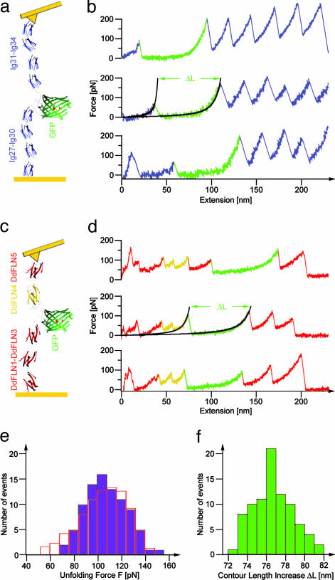
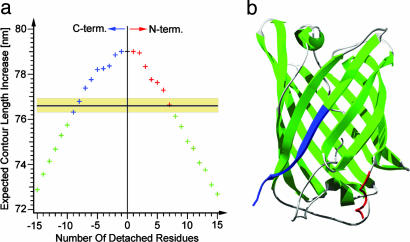
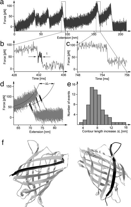
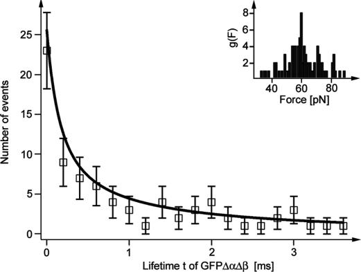
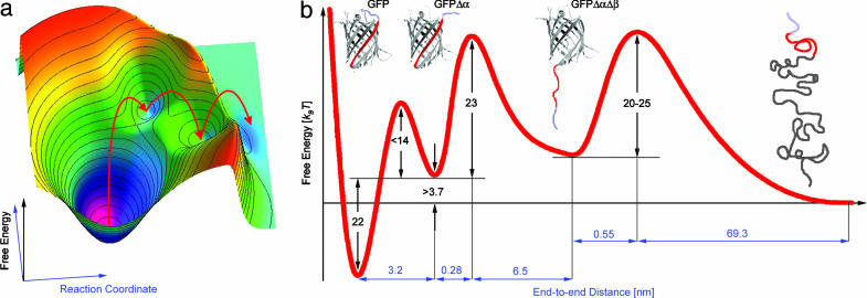
We use single-molecule force spectroscopy to drive single GFP molecules from the native state through their complex energy landscape into the completely unfolded state. Unlike many smaller proteins, mechanical GFP unfolding proceeds by means of two subsequent intermediate states. The transition from the native state to the first intermediate state occurs near thermal equilibrium at approximately 35 pN and is characterized by detachment of a seven-residue N-terminal alpha-helix from the beta barrel. We measure the equilibrium free energy cost associated with this transition as 22 k(B)T. Detachment of this small alpha-helix completely destabilizes GFP thermodynamically even though the beta-barrel is still intact and can bear load. Mechanical stability of the protein on the millisecond timescale, however, is determined by the activation barrier of unfolding the beta-barrel out of this thermodynamically unstable intermediate state. High bandwidth, time-resolved measurements of the cantilever relaxation phase upon unfolding of the beta-barrel revealed a second metastable mechanical intermediate with one complete beta-strand detached from the barrel. Quantitative analysis of force distributions and lifetimes lead to a detailed picture of the complex mechanical unfolding pathway through a rough energy landscape.


Figures

Fig. 1.
Fig. 1.
Single-domain force spectroscopy of GFP. (a) Scheme of Ig8-GFP chimera protein, stretched between a gold surface and a gold-coated cantilever tip. (b) Three typical force-extension traces measured with Ig8-GFP. GFP unfolding and the subsequent stretching phase of the unfolded polypeptide now lengthened by ΔL is marked in green. Black lines show WLC fits by using a persistence length, p, of 0.5 nm and contour lengths L and L + ΔL. (c) Scheme of DdFLN1-5-GFP chimera protein. (d) Three typical force-extension traces measured with DdFLN1-5-GFP. The GFP unfolding pattern is marked in green, and the double peak indicating DdFLN domain 4 unfolding (5) is marked in yellow. Black lines show WLC fits as in b. (e) Distribution of GFPΔα unfolding forces F (see Materials and Methods) obtained from measurements with DdFLN1-5-GFP. Open red bars show results from a Monte Carlo unfolding kinetics simulation of GFPΔα with τ0 = 14 s and Δx = 0.28 nm. (f) Distribution of the contour length increase, ΔL, because of GFPΔα unfolding as measured by WLC fits to DdFLN1-5-GFP traces (compare to d).
Fig. 3.
Fig. 3.
Detachment of the N-terminal α-helix. (a) Gray lines show detail of the region before GFPΔα unfolding of two Ig8-GFP force-extension traces. Black lines shows a fit with an elastically coupled two-state model (for fit parameters see text) (b) Gray line shows detail of a DdFLN1-5-GFP force-extension trace. Peak I corresponds to the unfolding of a DdFLN domain. Peak II corresponds to the unfolding of DdFLN domain 4. Peak III corresponds to the unfolding of GFPΔα. Black lines show a fit with an elastically coupled two-state model.
Fig. 2.
Fig. 2.
Contour length mapping. (a) Expected contour length increase because of unfolding of GFP as a function of the number of residues detached from either the N or C terminus from the native Cycle3-GFP structure (Protein Data Bank ID code 1B9C) (22). The black line with the shaded yellow region marks the measured contour length increase of 76.6 ± 0.3 nm. (b) Cycle3-GFP crystal structure (22). The regions marked in blue and red correspond to the C-terminal and N-terminal solution of the calculation in a.
Fig. 4.
Fig. 4.
Another short-lived intermediate. (a) DdFLN1-5-GFP force-extension trace measured at 10 kHz bandwidth with an Olympus type A Bio-Lever (resonance in water at 8.5 kHz). (b) Detail of the time course of the cantilever relaxation phase after unfolding of GFPΔα. The region marked in black indicates the presence of a short-lived intermediate state. (c) Detail of the time course of the cantilever relaxation phase after unfolding of a DdFLN domain. No deviations from normal damped oscillator relaxation were detected. (d) Six force-extension traces measured with DdFLN1-5-GFP and superimposed in the region of GFPΔα unfolding. The regions marked in black indicate the short-lived intermediate. All six intermediate levels fall on a WLC curve with a contour length increased by 6.8 nm. (e) Distribution of the contour length increase, ΔL, determined by WLC fits as shown in d. (f) Two possible structures of GFPΔαΔβ (light gray). Marked in dark gray are residues that unfold from the GFP barrel in the transition of GFPΔα to GFPΔαΔβ. (Left) A full β-strand detaches from the N terminus, and the folded GFP core comprises residues 25-230. (Right) A full β-strand detaches from the C terminus, and the folded GFP core comprises residues 11-209.
Fig. 5.
Fig. 5.
Lifetime distribution. Open squares indicate the distribution of measured lifetimes of GFPΔαΔβ. The black solid line shows a fit with a two-state model with Δx = 0.55 nm and τ0 = 10 s (see Materials and Methods). (Inset) Distribution of forces acting on GFPΔαΔβ.
Fig. 6.
Fig. 6.
Free energy surface. (a) Cartoon of the multidimensional energy landscape of GFP. The red arrows indicate the course of the mechanical unfolding pathway. (b) Projection of the energy landscape along the unfolding pathway onto one reaction coordinate.

References

    1. Clausen-Schaumann, H., Seitz, M., Krautbauer, R. & Gaub, H. E. (2000) Curr. Opin. Chem. Biol. 4, 524-530. - PubMed
    1. Best, R. B. & Clarke, J. (2002) Chem. Commun. 183-192. - PubMed
    1. Fisher, T. E., Marszalek, P. E. & Fernandez, J. M. (2000) Nat. Struct. Biol. 7, 719-724. - PubMed
    1. Li, H., Linke, W. A., Oberhauser, A. F., Carrion-Vazquez, M., Kerkvliet, J. G., Lu, H., Marszalek, P. E. & Fernandez, J. M. (2002) Nature 418, 998-1002. - PubMed
    1. Schwaiger, I., Kardinal, A., Schleicher, M., Noegel, A. A. & Rief, M. (2004) Nat. Struct. Mol. Biol. 11, 81-85. - PubMed

Publication types

Substances

LinkOut - more resources