Review
. 2005 Jan;6(1):40-6.
doi: 10.1002/cbic.200400274.

Chemical approaches to controlling intracellular protein degradation

Affiliations
Review

Chemical approaches to controlling intracellular protein degradation

John S Schneekloth Jr et al. Chembiochem. 2005 Jan.
No abstract available


Figures

Scheme 1
Scheme 1
Mechanism of RNA interference (RNAi). Double-stranded RNA is recognized by the Dicer enzyme and cleaved into 22-nucleotide siRNA fragments. These fragments are then separated by RISC, which associates with the antisense mRNA for the gene of interest to make an active complex. The RISC: mRNA complex cleaves the mRNA, resulting in gene silencing.
Scheme 2
Scheme 2
Chimeric F-box approach to biochemical protein degradation. In the wild-type E3 ligase, the F box recognizes the target protein, which is then ubiquitinated and ultimately degraded. If a binding domain for a target protein is engineered into the F box, a protein that is normally stable may be artificially ubiquitinated and degraded. E3=E3 ubiquitin ligase complex; Ub=ubiquitin; F=F-box-containing protein; BP=binding protein, recognition domain for the target protein.
Scheme 3
Scheme 3
Function of a PROTAC molecule. The PROTAC molecule induces complexation between the target protein and the E3 ubiquitin ligase. Once in a complex, the target protein is ubiquitinated and degraded. This approach requires no biochemical manipulation of the E3 ligase.
Scheme 4
Scheme 4
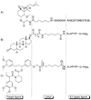
Structures of PROTAC molecules. A) fumagillin-based PROTAC. B) DHT-based PROTAC. C) AP21998-based PROTAC. Each PROTAC contains a target ligand, a linker, and an E3 ligase-recognition domain. Polyarginine tags are included for cell permeability. S*=phosphorylated serine.
Scheme 5
Scheme 5
Use of a PROTAC molecule to degrade a fluorescent protein. HEK 293 cells stably expressing GFP–androgen receptor were treated with 50 µm DHT–HIF-(Arg)8 PROTAC. After 1 h, the PROTAC induces ubiquitination and degradation of GFP–androgen receptor; this results in loss of fluorescence.

References

    1. Harvey DM, Caskey CT. Curr. Opin. Chem. Biol. 1998;2:512. - PubMed
    1. Dwek MV, Brooks SA. Curr. Cancer Drug Targets. 2004;4:425. - PubMed
    1. Bishop AC, Buzko O, Shokat KM. Trends Cell. Biol. 2001;11:167. - PubMed
    1. Shokat K, Velleca M. Drug Discovery Today. 2002;7:872. - PubMed
    1. Adams J. Curr. Opin. Oncol. 2002;14:628. - PubMed

Publication types

LinkOut - more resources