

Bovine cysticercosis: preliminary observations on the immunohistochemical detection of Taenia saginata antigens in lymph nodes of an experimentally infected calf
- PMID: 15532887
- PMCID: PMC545992
Bovine cysticercosis: preliminary observations on the immunohistochemical detection of Taenia saginata antigens in lymph nodes of an experimentally infected calf
Abstract
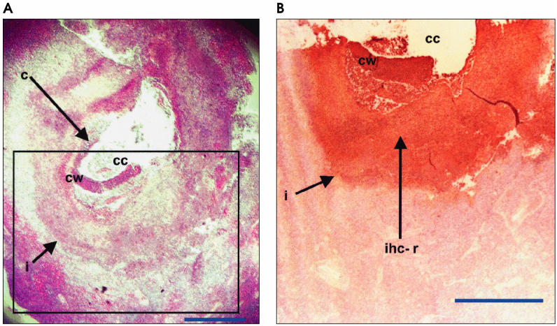
A newly developed immunohistochemical test was used for the first time to demonstrate the presence of Taenia saginata (Cysticercus bovis) antigens in the lymph nodes of a heifer calf experimentally inoculated with Taenia saginata eggs. The new test should aid in the differential diagnosis of eosinophilic lymphadenitis in cattle.
Résumé — Cysticercose bovine : observation préliminaire sur la détection immunohistochimique d’antigènes de Taenia saginata dans les ganglions lymphatiques d’un veau infecté expérimentalement. Un test immunohistochimique récemment mis au point a été utilisé pour la première fois afin de démontrer la présence d’antigènes de Taenia saginata (Cysticercus bovis) dans les ganglions lymphatiques d’une jeune génisse inoculée expérimentalement avec des œufs de Taenia saginata. Le nouveau test devrait aider à raffiner le diagnostic différentiel avec la lymphadénite eosinophilique chez les bovins.
(Traduit par Docteur André Blouin)
Figures

References
-
- Pawlowski ZS, Murrell KD. Taeniasis and cysticercosis. In: Hui YH, Sattar SA, Murrell KD, Nip W-K, Stanfield PS, eds. Foodborne Disease Handbook, 2nd ed. New York: Marcel Dekker, 2001:217–227.
-
- Gracey JF, Collins DS. Meat Hygiene, 9th ed. London: Ballière Tindall, 1992:307–351.
-
- Heath DD. The migration of oncospheres of Taenia pisiformis, T serialis and Echinoccocus granulosus within the intermediate host. Int J Parasitol. 1971;1:145–152. - PubMed
-
- Koudela K, Trefný D. The causality of Cysticercus bovis location in the heart and in the skeletal muscles of cattle. Helminthologia. 1973;14:163–169.
-
- Štěrba J, Dyková I, Machnicka B. Tissue reaction in the heart of cattle with a spontaneous and artificial Cysticercus bovis infection. Folia Parasitol (Praha) 1979;26:27–33. - PubMed
Publication types
MeSH terms
Substances
LinkOut - more resources
Full Text Sources