

Isoform-specific expression of the Coxsackie and adenovirus receptor (CAR) in neuromuscular junction and cardiac intercalated discs
- PMID: 15533241
- PMCID: PMC533869
- DOI: 10.1186/1471-2121-5-42
Isoform-specific expression of the Coxsackie and adenovirus receptor (CAR) in neuromuscular junction and cardiac intercalated discs
Abstract
Background: The Coxsackie and adenovirus receptor (CAR) has a restricted expression pattern in the adult. In skeletal muscle, although CAR is expressed in immature fibers, its transcript levels are barely detectable in mature muscle. This is in contrast to the robust expression observed in the heart. However, both heart and skeletal muscle are susceptible to infection with the Coxsackie B virus which utilizes primarily CAR for cellular internalization. The specific point of viral entry in skeletal and heart muscle remains unknown.
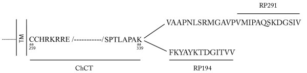
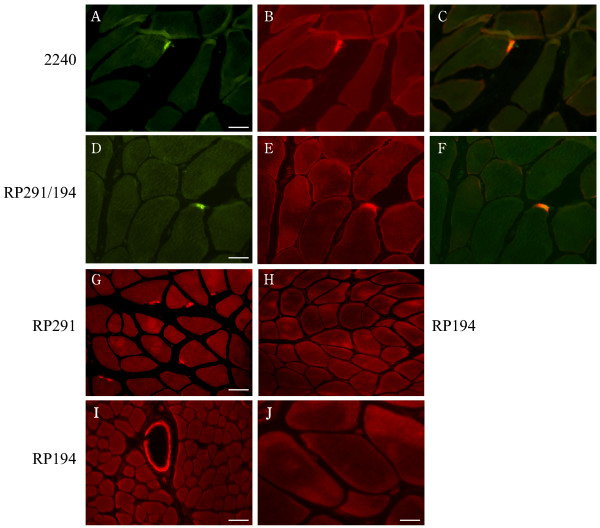
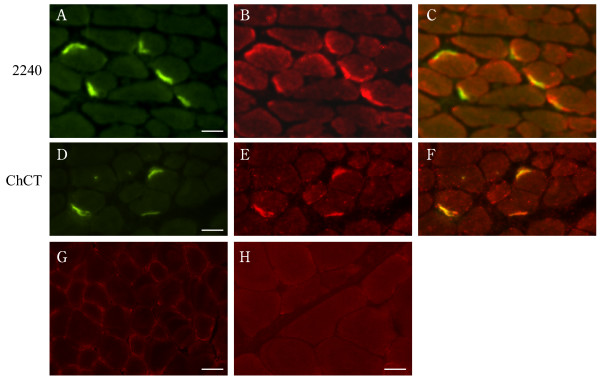
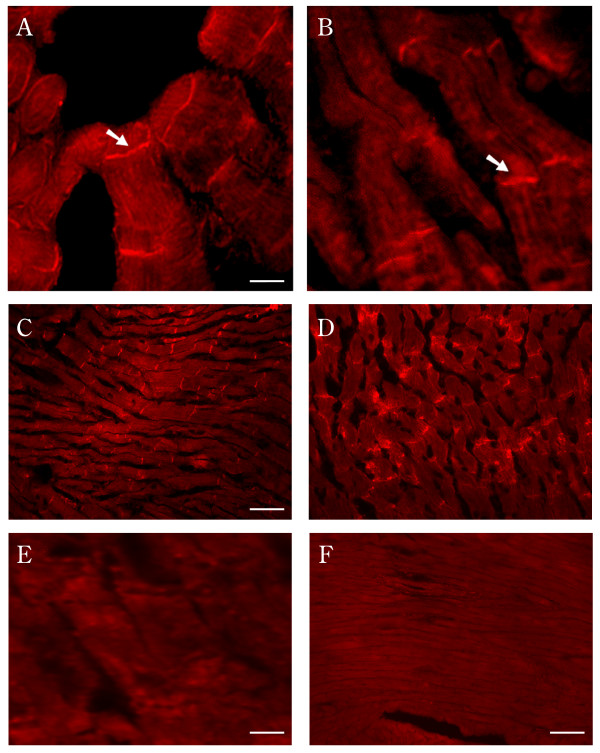
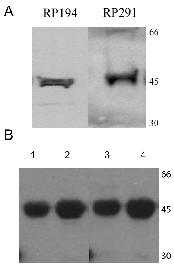
Results: Using antibodies directed against the extracellular and the cytoplasmic domains of CAR, we show CAR in normal human and mouse skeletal muscle to be a novel component of the neuromuscular junction. In cardiac muscle, CAR immunoreactivity is observed at the level of intercalated discs. We demonstrate a single isoform of CAR to be expressed exclusively at the human neuromuscular junction whereas both predominant CAR isoforms are expressed at the intercalated discs of non-diseased human heart.
Conclusion: The localization of CAR to these important junctional complexes suggests that CAR may play both a structural and a regulatory role in skeletal and cardiac muscle, and that these complexes may serve as a point of entry for Coxsackie B virus.
Figures
Similar articles
-
Localization of coxsackie virus and adenovirus receptor (CAR) in normal and regenerating human muscle.Neuromuscul Disord. 2005 Aug;15(8):541-8. doi: 10.1016/j.nmd.2005.05.007. Neuromuscul Disord. 2005. PMID: 16014330
-
Expression of the primary coxsackie and adenovirus receptor is downregulated during skeletal muscle maturation and limits the efficacy of adenovirus-mediated gene delivery to muscle cells.Hum Gene Ther. 1999 Apr 10;10(6):1009-19. doi: 10.1089/10430349950018409. Hum Gene Ther. 1999. PMID: 10223734
-
Spatial and temporal expression of the beta1D integrin during mouse development.Dev Dyn. 1997 Dec;210(4):472-86. doi: 10.1002/(SICI)1097-0177(199712)210:4<472::AID-AJA10>3.0.CO;2-9. Dev Dyn. 1997. PMID: 9415431
-
CAR: a virus receptor within the tight junction.Adv Drug Deliv Rev. 2005 Apr 25;57(6):869-82. doi: 10.1016/j.addr.2005.01.007. Adv Drug Deliv Rev. 2005. PMID: 15820557 Review.
-
Highly variable expression of virus receptors in the human cardiovascular system. Implications for cardiotropic viral infections and gene therapy.Z Kardiol. 2002 Dec;91(12):978-91. doi: 10.1007/s00392-002-0862-7. Z Kardiol. 2002. PMID: 12490988 Review.
Cited by
-
Effect of lovastatin on coxsackievirus B3 infection in human endothelial cells.Inflamm Res. 2014 Apr;63(4):267-76. doi: 10.1007/s00011-013-0695-z. Epub 2013 Dec 8. Inflamm Res. 2014. PMID: 24316867
-
Adenovirus transduction: More complicated than receptor expression.Virology. 2017 Feb;502:144-151. doi: 10.1016/j.virol.2016.12.020. Epub 2016 Dec 31. Virology. 2017. PMID: 28049062 Free PMC article.
-
Coxsackie and adenovirus receptor is a modifier of cardiac conduction and arrhythmia vulnerability in the setting of myocardial ischemia.J Am Coll Cardiol. 2014 Feb 18;63(6):549-59. doi: 10.1016/j.jacc.2013.10.062. Epub 2013 Nov 27. J Am Coll Cardiol. 2014. PMID: 24291282 Free PMC article.
-
The IgCAM CAR Regulates Gap Junction-Mediated Coupling on Embryonic Cardiomyocytes and Affects Their Beating Frequency.Life (Basel). 2022 Dec 21;13(1):14. doi: 10.3390/life13010014. Life (Basel). 2022. PMID: 36675963 Free PMC article.
-
The coxsackievirus-adenovirus receptor reveals complex homophilic and heterophilic interactions on neural cells.J Neurosci. 2010 Feb 24;30(8):2897-910. doi: 10.1523/JNEUROSCI.5725-09.2010. J Neurosci. 2010. PMID: 20181587 Free PMC article.
References
-
- Martino TA, Petric M, Weingartl H, Bergelson JM, Opavsky MA, Richardson CD, Modlin JF, Finberg RW, Kain KC, Willis N, Gauntt CJ, Liu PP. The coxsackie-adenovirus receptor (CAR) is used by reference strains and clinical isolates representing all six serotypes of coxsackievirus group B and by swine vesicular disease virus. Virology. 2000;271:99–108. doi: 10.1006/viro.2000.0324. - DOI - PubMed
Publication types
MeSH terms
Substances
LinkOut - more resources
Full Text Sources
Other Literature Sources
Molecular Biology Databases
