. 2004 Nov 8;167(3):531-43.
doi: 10.1083/jcb.200408165.

Recycling endosomes can serve as intermediates during transport from the Golgi to the plasma membrane of MDCK cells

Affiliations

Recycling endosomes can serve as intermediates during transport from the Golgi to the plasma membrane of MDCK cells

Agnes Lee Ang et al. J Cell Biol. .

Abstract

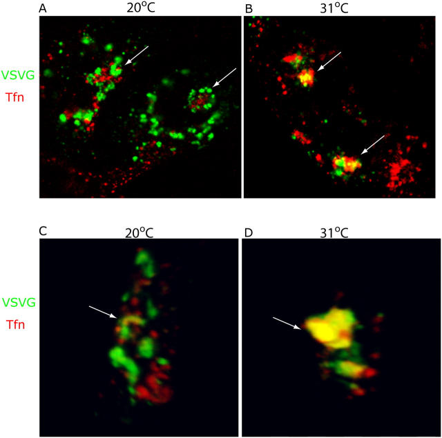
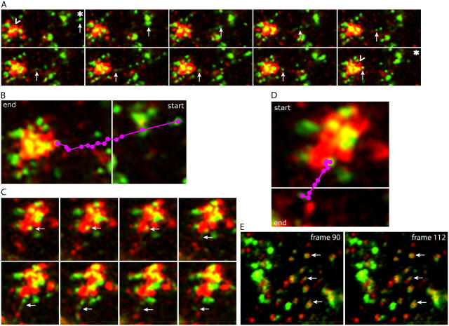
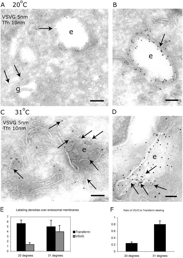
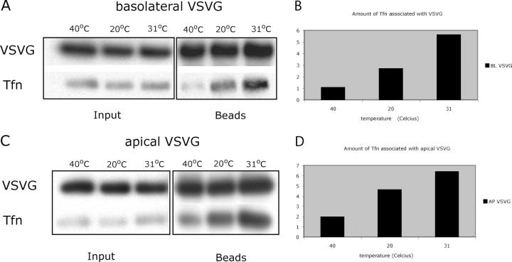
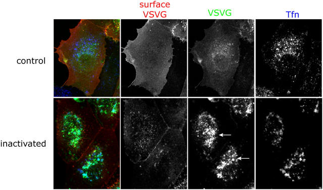
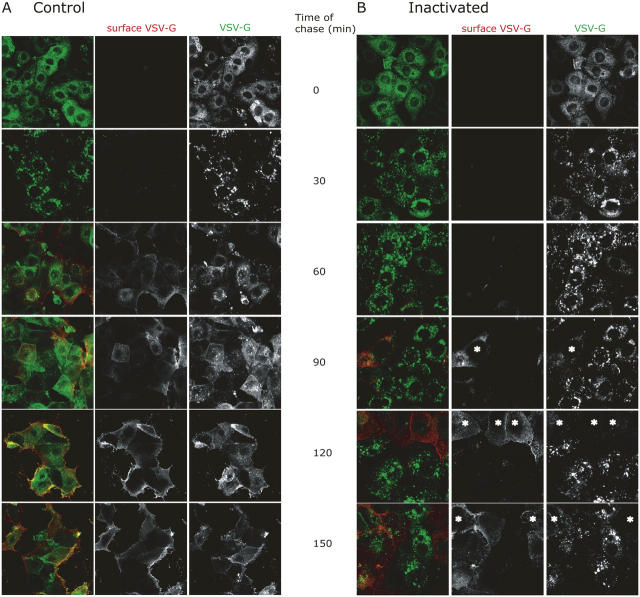
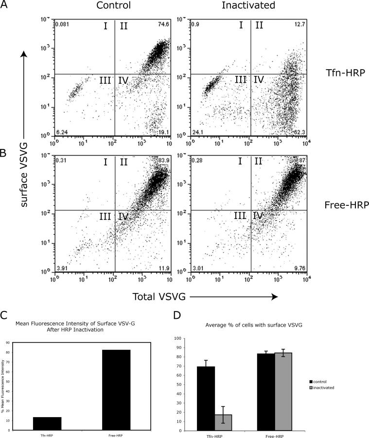
The AP-1B clathrin adaptor complex is responsible for the polarized transport of many basolateral membrane proteins in epithelial cells. Localization of AP-1B to recycling endosomes (REs) along with other components (exocyst subunits and Rab8) involved in AP-1B-dependent transport suggested that RE might be an intermediate between the Golgi and the plasma membrane. Although the involvement of endosomes in the secretory pathway has long been suspected, we now present direct evidence using four independent methods that REs play a role in basolateral transport in MDCK cells. Newly synthesized AP-1B-dependent cargo, vesicular stomatitis virus glycoprotein G (VSV-G), was found by video microscopy, immunoelectron microscopy, and cell fractionation to enter transferrin-positive REs within a few minutes after exit from the trans-Golgi network. Although transient, RE entry appears essential because enzymatic inactivation of REs blocked VSV-G delivery to the cell surface. Because an apically targeted VSV-G mutant behaved similarly, these results suggest that REs not only serve as an intermediate but also as a common site for polarized sorting on the endocytic and secretory pathways.


Figures

Figure 1.
Figure 1.
VSV-G localizes to Tfn-positive membranes after exit from the Golgi complex. (A) MDCKT cells were infected with ts045 VSV-G-YFP and induced to express transferrin receptor (TfnR). Cells were incubated 2 h at 20°C (last hour in media plus CHX) to accumulate a synchronous pulse of VSV-G (green) at the TGN, and allowed to take up Alexa 546-Tfn (red) into REs. Cells were fixed and imaged. Arrows, REs. (B) Cells in A were released from the TGN block by incubation at 31°C in media plus CHX for 10 min, and then fixed and imaged. Arrows denote colocalization of VSV-G and Tfn (yellow). (C and D) Three-dimensional reconstruction of confocal serial sections, X-Y plane and sagittal section through REs (arrow) of representative cells in A and B.
Figure 2.
Figure 2.
VSV-G is transported directly to and from REs as visualized by time-lapse microscopy. See Online supplemental material for original video images, available at http://www.jcb.org/cgi/content/full/jcb.200408165/DC1. (A) Individual frames from a movie of cells as prepared in Fig. 1 that were imaged immediately upon release of the TGN block by incubation at 31°C. Images were taken every 7 s for 30 min. Frame sequence illustrates the entry of VSV-G (green structures denoted by arrows; the asterisk marks starting point) into Tfn+ (red) RE structures. Note the increase of VSV-G in REs over the time course of these images, indicated by the increase in yellow (noted by “carrots” in the first and last frames). (B) Purple dots denote the path followed by VSV-G from the first frame (start) to the last frame in the sequence illustrated in A (end). (C) Sequence of VSV-G exit from REs, showing the apparent generation of a green VSV-G tubule and resulting vesicles from a yellow structure contained within the red RE region (arrows). Time course is the same as in A. (D) Purple dots denote the path of VSV-G exit from first frame (start) to last frame (end) in the sequence illustrated in panel C. (E) Image of cell periphery from a cell prepared as in A. Images were taken every 7 s. Arrows denote colocalization of VSV-G with Tfn, an association that was maintained >2.5 min, suggesting that the VSV-G and Tfn were contained within the same structure or reflected two distinct but tethered structures.
Figure 3.
Figure 3.
VSV-G and Tfn colocalize in endosomes by immuno-EM. (A and B) Immuno-EM of MDCKT cells expressing VSV-G-YFP and having endocytosed Alexa 488-Tfn after 2-h incubation at 20°C. VSV-G (5 nm gold, arrows) was localized to the Golgi complex, peripheral vesicles, and, occasionally, on endosomes (arrow on “e”). Tfn (10 nm gold) was localized to endosomes (e). Bars: (A) 200 nm; (B) 100 nm. (C and D) Immuno-EM of MDCKT cells released from the 20°C TGN block by incubation at 31°C for 10 min. VSV-G (5 nm gold, arrows) and Tfn (10 nm gold) were localized to the same endosomal compartments (e). Bars, 100 nm. (E) Labeling density of VSV-G on Tfn+ endosomes. Density of VSV-G labeling increases fourfold during 31°C incubation. (F) Ratio of number of gold particles labeling VSV-G in endosomes over the number of gold particles labeling Tfn. Error bars represent the SD of labeling density from three grids.
Figure 4.
Figure 4.
Immunoisolated VSV-G membranes from MDCKT are associated with Tfn after exit from the TGN. (A) Representative Western blot of immunoisolated basolateral VSV-G-YFP containing membranes from VSV-G-YFP–infected MDCKT cells incubated with Alexa 488-Tfn during a 40°C ER block, 20°C TGN block, and 10-min chase at 31°C. Immunoprecipitation of VSV-G was performed using magnetic beads coupled to anti-GFP antibody (beads lanes) and probed for VSV-G (VSV-G row). VSV-G purified membranes were also probed for the presence of Alexa 488-Tfn using anti–Alexa 488 antibody (Tfn lane). This is a representative blot from four experiments. (C) Same as in A but apical mutant VSV-G-G3-YFP was used; representative blot from three experiments. (B and D) Quantification of Tfn associated with VSV-G under the three temperature conditions from A and B.
Figure 5.
Figure 5.
Inactivation of REs causes accumulation of VSV-G in the perinuclear region. MDCKT cells were infected to express VSV-G-YFP (third column, green) and incubated overnight at 40°C. Tfn-HRP (third column, blue) was internalized for 45 min at 40°C and was chased into REs by incubating cells in media without Tfn-HRP for 15 min at 40°C. Control cells were subject to only DAB while the samples were exposed to DAB and H2O2 for 1 h in the dark. Cells were washed in warm media and incubated at 31°C for 1.5 h in media/CHX to release VSV-G from the ER. Cells were washed in PBScmf, trypsinized, fixed, and processed for immunofluorescence. Cell surface VSV-G labeling (second column, red) using an antibody, TKG, against the ectodomain of VSV-G was performed on nonpermeabilized cells before permeabilization and internal labeling for HRP. Arrows denote accumulation of intracellular VSV-G in the perinuclear region.
Figure 6.
Figure 6.
Inactivation of RE caused an inhibition of VSV-G transport in the secretory pathway. MDCKT cells were prepared identically as in Fig. 5 but processed for immunofluorescence every 30 min up to 2.5 h total time of chase at 31°C. Peroxide was not present in control cells (A), whereas samples whose REs were inactivated were subject to peroxide (B). Cells in B that apparently escaped the DAB inactivation at long times of chase are marked by asterisks. Red, surface VSV-G (TKG staining); green, total VSV-G fluorescence.
Figure 7.
Figure 7.
Inactivation of membranes containing Tfn-HRP, but not free-HRP, inhibited the cell surface arrival of VSV-G. (A) Representative dot plot of flow cytometry results from cells prepared as in Fig. 5 (Tfn-HRP). Levels of surface VSV-G, performed on nonpermeabilized cells using the TKG antibody, were quantified on the y-axis whereas total VSV-G, as monitored by YFP fluorescence, was quantified on the x-axis. Cells in quadrant I were positive for only surface VSV-G, cells in quadrant II were positive for both surface and total VSV-G, cells in quadrant III were negative for both markers, and cells in quadrant IV were negative for surface VSV-G and positive for total (i.e., intracellular) VSV-G. Numbers in corners represent percentage of cells in that quadrant. (B) MDCKT cells expressing VSV-G-YFP were incubated with free-HRP followed by chase in HRP-free media to load HRP into lysosomes (Free-HRP). Control cells were incubated with DAB alone while the “inactivated” set was incubated with DAB plus H2O2 for 1 h in the dark. (C) Percentage MFI of surface VSV-G was measured in cells that were positive for total VSV-G (all cells in quadrants II and IV) and had Tfn-HRP– or free-HRP–containing compartments inactivated. MFI was normalized based on levels in control cells. (D) Average percentage of cells based on three experiments performed in triplicate with surface VSV-G after an uptake of Tfn-HRP or free-HRP under control (black) or inactivation (gray) conditions. Error bars represent the SD from cells with surface VSV-G from three different experiments.

References

    1. Ang, A.L., H. Fölsch, U.M. Koivisto, M. Pypaert, and I. Mellman. 2003. The Rab8 GTPase selectively regulates AP-1B–dependent basolateral transport in polarized Madin-Darby canine kidney cells. J. Cell Biol. 163:339–350. - PMC - PubMed
    1. Bartles, J.R., H.M. Feracci, B. Stieger, and A.L. Hubbard. 1987. Biogenesis of the rat hepatocyte plasma membrane in vivo: comparison of the pathways taken by apical and basolateral proteins using subcellular fractionation. J. Cell Biol. 105:1241–1251. - PMC - PubMed
    1. Bonifacino, J.S., and L.M. Traub. 2003. Signals for sorting of transmembrane proteins to endosomes and lysosomes. Annu. Rev. Biochem. 72:395–447. - PubMed
    1. Casanova, J.E., X. Wang, R. Kumar, S.G. Bhartur, J. Navarre, J.E. Woodrum, Y. Altschuler, G.S. Ray, and J.R. Goldenring. 1999. Association of Rab25 and Rab11a with the apical recycling system of polarized Madin-Darby canine kidney cells. Mol. Biol. Cell. 10:47–61. - PMC - PubMed
    1. Chen, W., Y. Feng, D. Chen, and A. Wandinger-Ness. 1998. Rab11 is required for trans-golgi network-to-plasma membrane transport and a preferential target for GDP dissociation inhibitor. Mol. Biol. Cell. 9:3241–3257. - PMC - PubMed

Publication types