Comparative Study
. 2004 Nov 16;101(46):16268-73.
doi: 10.1073/pnas.0407359101. Epub 2004 Nov 8.

Crystal structure of a human CD3-epsilon/delta dimer in complex with a UCHT1 single-chain antibody fragment

Affiliations
Comparative Study

Crystal structure of a human CD3-epsilon/delta dimer in complex with a UCHT1 single-chain antibody fragment

Kelly L Arnett et al. Proc Natl Acad Sci U S A. .

Abstract

The alpha/beta T cell receptor complex transmits signals from MHC/peptide antigens through a set of constitutively associated signaling molecules, including CD3-epsilon/gamma and CD3-epsilon/delta. We report the crystal structure at 1.9-A resolution of a complex between a human CD3-epsilon/delta ectodomain heterodimer and a single-chain fragment of the UCHT1 antibody. CD3-epsilon/delta and CD3-epsilon/gamma share a conserved interface between the Ig-fold ectodomains, with parallel packing of the two G strands. CD3-delta has a more electronegative surface and a more compact Ig fold than CD3-gamma; thus, the two CD3 heterodimers have distinctly different molecular surfaces. The UCHT1 antibody binds near an acidic region of CD3-epsilon opposite the dimer interface, occluding this region from direct interaction with the TCR. This immunodominant epitope may be a uniquely accessible surface in the TCR/CD3 complex, because there is overlap between the binding site of the UCHT1 and OKT3 antibodies. Determination of the CD3-epsilon/delta structure completes the set of TCR/CD3 globular ectodomains and contributes information about exposed CD3 surfaces.


Figures
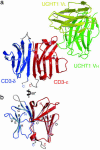
Fig. 1.
Fig. 1.
Structure of the CD3-ε/δ/UCHT1-scFv complex and topology of the CD3-ε/δ dimer. (a) Ribbon diagram illustrating CD3-ε (red), CD3-δ (blue), UCHT1 heavy chain variable domain (green), and UCHT1 light chain variable domain (yellow). The eight CD3-ε strands and seven CD3-δ strands are labeled A-G by using standard nomenclature for I-set and C1-set Ig folds, respectively. Residues modeled using weak density or density from the second data set are in gray. Glycosylation sites of CD3-δ at Asn-17 and Asn-53 (gray sticks) are marked, although no glycans are present in this bacterially expressed protein. Disulfide bonds between B and F strands are in orange. (b) Sixty-degree rotation of the CD3-ε/δ dimer around the axis formed by G-strand pairing at the dimer interface.
Fig. 2.
Fig. 2.
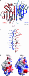
Comparison of human CD3-ε/δ and human CD3-ε/γ. (a) Overlay of CD3-ε/δ (red/blue) and CD3-ε/γ (gray), created by superimposing the CD3-ε subunits. (b) Backbone and side-chain detail of G-strand pairing of CD3 dimers. Main-chain hydrogen bonds between CD3-ε and CD3-δ are shown as dotted cyan lines. Residues absolutely conserved between CD3-γ and CD3-δ or residues conserved among mammalian CD3-ε are labeled with**; those that conserve similarity are labeled with*. Human CD3-γ residue differences are indicated in parentheses. (c) Positive and negative electrostatic surface potentials (blue and red, respectively) of CD3-ε/δ and CD3-ε/γ dimers. The view is a 90° rotation from the one in a, showing a side view of CD3-δ and CD3-γ on the left and right, respectively. In contrast to CD3-γ, which has an electropositive surface, both CD3-ε and CD3-δ are electronegative.
Fig. 3.
Fig. 3.
Molecular surface representations of CD3-ε/δ conservation. Residues conserved among mammalian CD3-ε molecules (red), residues conserved between CD3-δ and CD3-γ (green), and residues conserved among CD3-δ molecules but not found in CD3-γ (blue) are colored. (a) “Front” face of CD3-ε/δ oriented such that the C-terminal residues of CD3-ε and CD3-δ are at the bottom and CD3-ε and CD3-δ subunits are side-to-side, as in Fig. 2a. (b) Side of CD3-δ. (c) “Back” face of CD3-ε/δ, as in Fig. 1 (a 180° rotation of a). (d) Side of CD3-ε. The front, back, and CD3-δ side of the dimer have clusters of conserved surface residues. The CD3-ε side of the dimer is devoid of conserved regions.
Fig. 4.
Fig. 4.
Molecular surface of CD3-ε/δ and UCHT1 contact residues. Positive and negative electrostatic surface potentials of CD3-ε/δ dimer are indicated on the translucent molecular surface in blue and red, respectively. CD3-ε/δ is oriented as in Fig. 1. CD3-ε and UCHT1 contact residues, which form hydrogen bonds or salt bridges, are indicated as ball-and-stick models. UCHT1 light chain (yellow) and heavy chain (green) bind exclusively to CD3-ε near an electronegative region on the side (indicated with an arrow), partially occluding it and burying a molecular surface of 1,789 Å2.
Fig. 5.
Fig. 5.
Conservation of TCR/CD3 extracellular domains and proposed model for TCR/CD3 complex. (a) Schematic representation of TCR and CD3 assembly. Conserved regions are shown as bold patches. Nonconserved regions and TCR variable domains are pale. Carbohydrate moieties are gray spheres. (b) A proposed model for the TCR/CD3 complex. Its principle features are (i) the complex is tight, because trimeric transmembrane contacts among TCR/CD3 components (α-ε-δ, β-ε-γ, and α-ζ-ζ) suggest a compact bundle, perhaps no wider than the TCR alone; (ii) to create these interactions, the CD3 dimer ectodomains lie angled to the membrane between it and the TCR globular domains; and (iii) CD3-ε/γ and CD3-ε/δ interact by their heterodimeric faces with asymmetric nonglycosylated TCR surfaces that are conserved among mammals. The lengths of TCR-β and TCR-α connecting peptides missing from known structures suggest that the TCR sits “above” the CD3 dimers. The requirement that TCR and CD3 TM domains interact, the shape of the membrane-proximal surface of the TCR, and the positions of conserved residues in the three heterodimers suggest that the paddle-shaped CD3 ectodomains lie at an angle relative to the membrane. The stoichiometry and interspecies promiscuity of TCR/CD3 interactions suggest that both CD3 dimers interact through their heterodimeric faces with the conserved nonglycosylated bottom and side of the TCR. This interaction mode would allow contacts to extend to the small patches of conserved residues on the sides of CD3-γ or CD3-δ. Glycosylation of these sides in some species precludes direct side-on binding with the TCR.

References

    1. Davis, M. M. & Bjorkman, P. J. (1988) Nature 334, 395-402. - PubMed
    1. Clevers, H., Alarcon, B., Wileman, T. & Terhorst, C. (1988) Annu. Rev. Immunol. 6, 629-662. - PubMed
    1. Letourneur, F. & Klausner, R. D. (1992) Science 255, 79-82. - PubMed
    1. Koning, F., Maloy, W. L. & Coligan, J. E. (1990) Eur. J. Immunol. 20, 299-305. - PubMed
    1. Manolios, N., Letourneur, F., Bonifacino, J. S. & Klausner, R. D. (1991) EMBO J. 10, 1643-1651. - PMC - PubMed

Publication types

Associated data