

Thyroid hormone administration enhances remyelination in chronic demyelinating inflammatory disease
- PMID: 15534218
- PMCID: PMC526198
- DOI: 10.1073/pnas.0407262101
Thyroid hormone administration enhances remyelination in chronic demyelinating inflammatory disease
Abstract
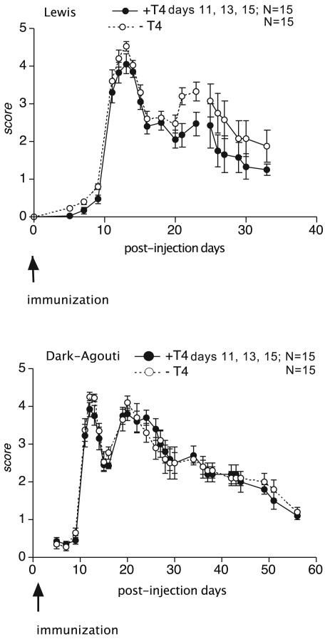
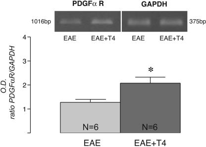
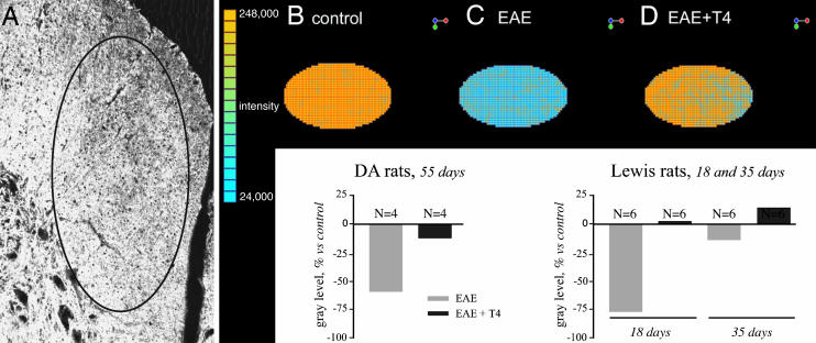
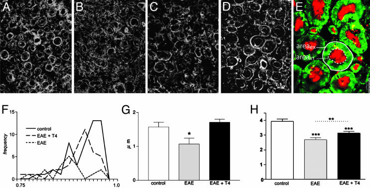
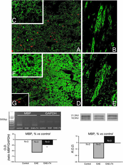
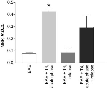
Chronic disabilities in multiple sclerosis are believed to be due to neuron damage and degeneration, which follow remyelination failure. Due to the presence of numerous oligodendrocyte precursors inside demyelination plaques, one reason for demyelination failure could be the inability of oligodendrocyte precursor cells to turn into myelinating oligodendrocytes. In this study, we show that thyroid hormone enhances and accelerates remyelination in an experimental model of chronic demyelination, i.e., experimental allergic encephalomyelitis in congenic female Dark Agouti rats immunized with complete guinea pig spinal cord. Thyroid hormone, when administered during the acute phase of the disease, increases expression of platelet-derived growth factor alpha receptor, restores normal levels of myelin basic protein mRNA and protein, and allows an early and morphologically competent reassembly of myelin sheaths. Moreover, thyroid hormone exerts a neuroprotective effect with respect to axonal pathology.
Figures
Similar articles
-
The neuregulin, glial growth factor 2, diminishes autoimmune demyelination and enhances remyelination in a chronic relapsing model for multiple sclerosis.Proc Natl Acad Sci U S A. 1998 Aug 18;95(17):10100-5. doi: 10.1073/pnas.95.17.10100. Proc Natl Acad Sci U S A. 1998. PMID: 9707607 Free PMC article.
-
Ellagic acid protects from myelin-associated sphingolipid loss in experimental autoimmune encephalomyelitis.Biochim Biophys Acta Mol Cell Biol Lipids. 2018 Sep;1863(9):958-967. doi: 10.1016/j.bbalip.2018.05.009. Epub 2018 May 21. Biochim Biophys Acta Mol Cell Biol Lipids. 2018. PMID: 29793057
-
Thyroid hormone activates oligodendrocyte precursors and increases a myelin-forming protein and NGF content in the spinal cord during experimental allergic encephalomyelitis.Proc Natl Acad Sci U S A. 2002 Mar 5;99(5):3258-63. doi: 10.1073/pnas.052704499. Epub 2002 Feb 26. Proc Natl Acad Sci U S A. 2002. PMID: 11867745 Free PMC article.
-
Nudging oligodendrocyte intrinsic signaling to remyelinate and repair: Estrogen receptor ligand effects.J Steroid Biochem Mol Biol. 2016 Jun;160:43-52. doi: 10.1016/j.jsbmb.2016.01.006. Epub 2016 Jan 14. J Steroid Biochem Mol Biol. 2016. PMID: 26776441 Free PMC article. Review.
-
Thyroid hormone and remyelination in adult central nervous system: a lesson from an inflammatory-demyelinating disease.Brain Res Brain Res Rev. 2005 Apr;48(2):339-46. doi: 10.1016/j.brainresrev.2004.12.022. Epub 2005 Jan 26. Brain Res Brain Res Rev. 2005. PMID: 15850672 Review.
Cited by
-
Effect of Mild Thyrotoxicosis on Performance and Brain Activations in a Working Memory Task.PLoS One. 2016 Aug 18;11(8):e0161552. doi: 10.1371/journal.pone.0161552. eCollection 2016. PLoS One. 2016. PMID: 27536945 Free PMC article. Clinical Trial.
-
Oligodendroglial Lineage Cells in Thyroid Hormone-Deprived Conditions.Stem Cells Int. 2019 Apr 30;2019:5496891. doi: 10.1155/2019/5496891. eCollection 2019. Stem Cells Int. 2019. PMID: 31182964 Free PMC article. Review.
-
Nuclear hormone receptors in demyelinating diseases.J Neuroendocrinol. 2022 Jul;34(7):e13171. doi: 10.1111/jne.13171. Epub 2022 Jun 22. J Neuroendocrinol. 2022. PMID: 35734821 Free PMC article. Review.
-
In vitro and in vivo pharmacological models to assess demyelination and remyelination.Neuropsychopharmacology. 2009 Jan;34(1):55-73. doi: 10.1038/npp.2008.145. Epub 2008 Sep 17. Neuropsychopharmacology. 2009. PMID: 18800062 Free PMC article. Review.
-
The role of thyroid hormones as inductors of oxidative stress and neurodegeneration.Oxid Med Cell Longev. 2013;2013:218145. doi: 10.1155/2013/218145. Epub 2013 Dec 9. Oxid Med Cell Longev. 2013. PMID: 24386502 Free PMC article. Review.
References
-
- Compston, A. & Coles, A. (2002) Lancet 359, 1221-1231. - PubMed
-
- Ferguson, B., Matyszak, M. K., Eiri, M. M. & Perry, V. H. (1997) Brain 120, 393-399. - PubMed
-
- De Stefano, N., Matthews, P. M., Fu, L., Narayanan, S., Stanley, J., Francis, G. S., Antel, J. P. & Arnold, D. L. (1998) Brain 121, 1469-1477. - PubMed
Publication types
MeSH terms
Substances
LinkOut - more resources
Full Text Sources
Other Literature Sources
Molecular Biology Databases
