. 2004 Nov 16;101(46):16339-44.
doi: 10.1073/pnas.0407416101. Epub 2004 Nov 8.

RGS9-2 modulates D2 dopamine receptor-mediated Ca2+ channel inhibition in rat striatal cholinergic interneurons

Affiliations

RGS9-2 modulates D2 dopamine receptor-mediated Ca2+ channel inhibition in rat striatal cholinergic interneurons

Theresa M Cabrera-Vera et al. Proc Natl Acad Sci U S A. .

Abstract

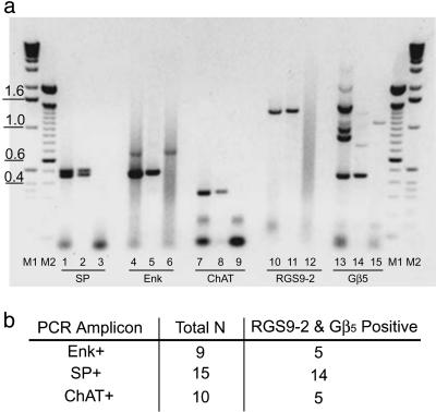
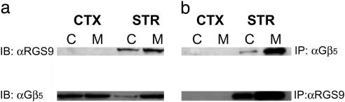
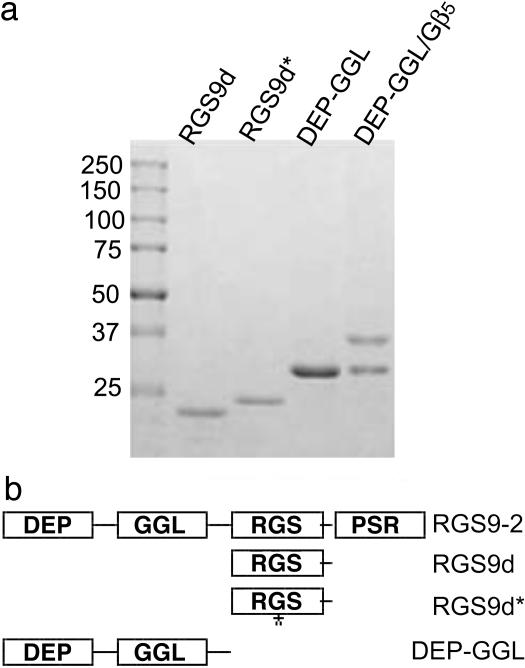
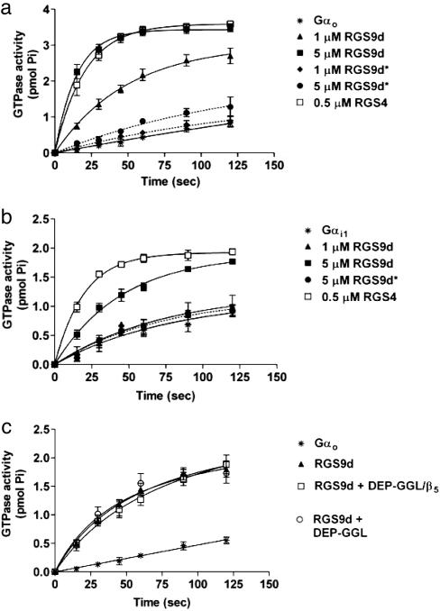
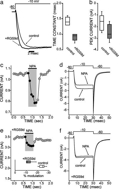
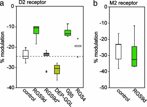
Regulator of G protein signaling (RGS) proteins negatively regulate receptor-mediated second messenger responses by enhancing the GTPase activity of Galpha subunits. We describe a receptor-specific role for an RGS protein at the level of an individual brain neuron. RGS9-2 and Gbeta(5) mRNA and protein complexes were detected in striatal cholinergic and gamma-aminobutyric acidergic neurons. Dialysis of cholinergic neurons with RGS9 constructs enhanced basal Ca(2+) channel currents and reduced D(2) dopamine receptor modulation of Cav2.2 channels. These constructs did not alter M(2) muscarinic receptor modulation of Cav2.2 currents in the same neuron. The noncatalytic DEP-GGL domain of RGS9 antagonized endogenous RGS9-2 activity, enhancing D(2) receptor modulation of Ca(2+) currents. In vitro, RGS9 constructs accelerated GTPase activity, in agreement with electrophysiological measurements, and did so more effectively at Go than Gi. These results implicate RGS9-2 as a specific regulator of dopamine receptor-mediated signaling in the striatum and identify a role for GAP activity modulation by the DEP-GGL domain.


Figures

Fig. 1.
Fig. 1.
RGS9-2 and Gβ5 mRNA are present in medium spiny neurons and in cholinergic interneurons within the striatum. (a) Representative ethidium bromide staining of RT-PCR products generated from either a single striatal neuron (lanes 2, 5, 8, 11, and 14) or a striatal cDNA-positive control preparation (lanes 1, 4, 7, 10, and 13). Lanes 3, 6, 9, 12, and 15 show the negative controls where no cDNA template was added to the PCR. Additional negative controls included the omission of reverse transcriptase (control for genomic DNA contamination of RNA samples). No PCRs were detected in these controls (data not shown). PCR fragments observed were of the expected sizes generated from rat gene-specific primers complementary to either substance P (doublet at 468 and 513 bp), enkephalin (477 bp), choline acetyltransferase (324 bp), RGS9-2 (1,300 bp), or Gβ5 (448 bp). M1 and M2 denote DNA standard markers. (b) Summary of single cell RT-PCR results indicating that RGS9-2 and Gβ5 are present in all neuronal subtypes examined. Total N is the total number of cells examined for each neuronal subtype. The number of cells positive for both RGS9-2 and Gβ5 mRNA are indicated to the right of the total.
Fig. 2.
Fig. 2.
RGS9-2/Gβ5 complexes coimmunoprecipitate from the striatum. (a) Western blot showing RGS9-2 detected in striatal homogenates (STR) but not in cortical preparations (CTX), whereas Gβ5 is present in both. RGS9-2 and Gβ5 are found in both cytosolic (C) and membrane (M) extracts. (b) RGS9-2 and Gβ5 coimmunoprecipitate from both membrane and cytosolic compartments of the striatum with either RGS9 or Gβ5 antibodies, but do not coimmunoprecipitate from cortical negative controls. Data shown are representative of three identical experiments conducted. IP, immunoprecipitating antibody; IB, antibody used to probe Western blot after immunoprecipitation.
Fig. 3.
Fig. 3.
Expression and purification of RGS9 constructs. (a) Coomassie-stained SDS/PAGE gel of N-terminally His6-tagged purified RGS9 constructs. Preparations were >90% pure. (b) Schematic diagram of RGS9 constructs. Domains are DEP (Dishevled/Egl10/Plextrin), GGL (G gamma-like), RGS (Regulator of G protein signaling), and PSR (proline/serine-rich).
Fig. 4.
Fig. 4.
RGS9 stimulates the intrinsic GTPase activity of Gαo and Gαi1 in a single-turnover assay. (a) RGS9d accelerates Go GTPase activity (intrinsic rate constant k = 0.002 min-1) in a concentration-dependent manner (1 μM RGS9d, k = 0.021; 5 μM RGS9d, k = 0.067 min-1). Introduction of the point mutation I363T within the RGS domain near a critical Gi1 contact site (RGS9d*) eliminates the GTPase activity of this construct (1 μM RGS9d, k = 0.003; 5 μM RGS9d, k = 0.007 min-1). (b) RGS9d accelerates Gi1 GTPase activity (intrinsic rate constant k = 0.01 min-1) with lower potency than that observed for Gαo. Also similar to Gαo, RGS9d* is devoid of GAP activity toward Gαi1 (5 μM RGS9d*, k = 0.012; 1 μM RGS9d, k = 0.014; 5 μM RGS9d, k = 0.022 min-1). (c) Neither DEP-GGL nor DEP-GGL/Gβ5 directly influences the GAP activity of RGS9d toward Gαo. Neither construct affects the GTPase activity of Gαo in the absence of RGS9d.
Fig. 5.
Fig. 5.
The RGS domain of RGS9 (RGS9d, 10 μM) alters Ca2+ current modulation in striatal cholinergic interneurons, both in the absence of agonist (a and b) and in the presence of D2-dopaminergic receptor stimulation (c-f). In the presence of a GPCR agonist, the Ca2+ current is modulated so that the peak current is decreased (c and d). Addition of RGS9d in the pipette decreases this Ca2+ current modulation (e and f). (a and b) In the absence of agonist, addition of the RGS domain in the pipette speeds up the activation kinetics of the current (a), and the current amplitude increases (b). Patch clamp recordings showing the onset kinetics of Ca2+ currents in control conditions and with RGS9d included in the patch pipette in two typical neurons. Stimulatory step from -80 mV to -10 mV. (b) The box plot summary of the current amplitude in control conditions and in the presence of RGS9d (n = 6, P < 0.05 Kruskall-Wallis ANOVA). For smaller sized groups of data, where means do not necessarily give a good measure of central tendency, medians and ranges are given with box plots. In these plots, the central bar of the box represents the median, and the edges are the interquartiles (technically, fourths). The bars are lines drawn to the most extreme points in the sample group that are not outliers (defined as points beyond interquartile ± 1.5 interquartile range). (c) Plot of the peak current evoked by a pulse from -80 to -10 mV as a function of time; application of the D2 agonist R(-)-propylnorapomorphine (NPA, 10 μM) is indicated by the bar above the trace. (d) Individual current traces of voltage-activated Ca2+ currents after a stimulatory voltage step from -80 mV to -10 mV, then back to -60 mV. This trace shows the reduction in the current amplitude by NPA in the same cell as in a. (e) Plot of the peak current shows the NPA modulation of the Ca2+ current in one cell loaded with RGS9d. (Inset) The box plot summary of the current modulation by NPA in control conditions (n = 5) and in the presence of RGS9d (n = 6, P < 0.05 Kruskal-Wallis ANOVA). (f) Current traces corresponding to the same cell as shown in c.
Fig. 6.
Fig. 6.
Summary of the effects of different domains of RGS9-2 on D2 dopaminergic and M2 muscarinic modulation of Ca2+ channels. (a) Under control conditions, D2 dopaminergic stimulation causes an ≈25% decrease in Cav2.2 Ca2+ channel currents as shown in Fig. 4a. Introduction of the RGS domain of RGS9 results in a 50% decrease in D2-dopaminergic modulation of Ca2+ channels, by increasing the rate of Gi turnoff (see also Fig. 4c). RGS4 has no significant effect, demonstrating that this effect is specific to RGS9. Application of an RGS9 construct that is catalytically inactive, RGS9d*, fails to alter the D2 receptor-mediated modulation of Ca2+ currents. Introduction of the DEP/GGL domain of RGS9 (10 μM) antagonizes endogenous RGS9-2 and increases the impact of D2 receptor activation on Ca2+ channel currents. Introduction of Gβ5 decreases the ability of dopamine to modulate Ca2+ current as well as RGS9d, suggesting stabilization or recruitment of endogenous RGS9-2 to the membrane. (b) RGS9d does not modulate M2 muscarinic receptor-mediated signaling to Ca2+ channels, thereby demonstrating receptor-specific regulation by RGS9d. The box plot summary (see Fig. 5) of the current amplitude in control conditions and in the presence of RGS9d (n = 6, P < 0.05, Kruskal-Wallis ANOVA) is shown.

References

    1. Martemyanov, K. A., Lishko, P. V., Calero, N., Keresztes, G., Sokolov, M., Strissel, K. J., Leskov, I. B., Hopp, J. A., Kolesnikov, A. V., Chen, C. K., et al. (2003) J. Neurosci. 23, 10175-10181. - PMC - PubMed
    1. Snow, B. E., Krumins, A. M., Brothers, G. M., Lee, S. F., Wall, M. A., Chung, S., Mangion, J., Arya, S., Gilman, A. G. & Siderovski, D. P. (1998) Proc. Natl. Acad. Sci. USA 95, 13307-13312. - PMC - PubMed
    1. Posner, B. A., Gilman, A. G. & Harris, B. A. (1999) J. Biol. Chem. 274, 31087-31093. - PubMed
    1. Snow, B. E., Betts, L., Mangion, J., Sondek, J. & Siderovski, D. P. (1999) Proc. Natl. Acad. Sci. USA 96, 6489-6494. - PMC - PubMed
    1. Liang, J. J., Chen, H. H., Jones, P. G. & Khawaja, X. Z. (2000) J. Neurosci. Res. 60, 58-64. - PubMed

Publication types

MeSH terms

LinkOut - more resources