. 2005 Jun 15;388(Pt 3):967-72.
doi: 10.1042/BJ20041066.

The membrane-anchored serine protease, TMPRSS2, activates PAR-2 in prostate cancer cells

Affiliations

The membrane-anchored serine protease, TMPRSS2, activates PAR-2 in prostate cancer cells

Susan Wilson et al. Biochem J. .

Abstract

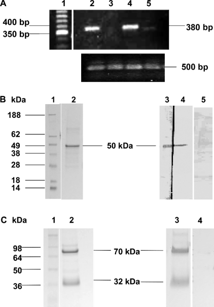
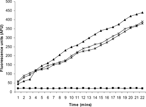
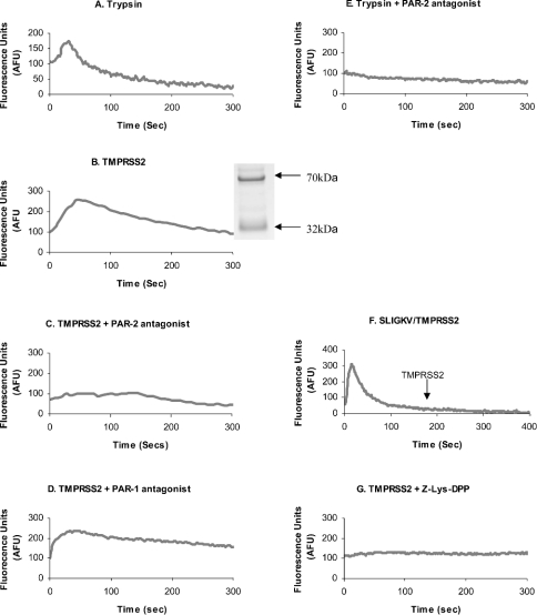
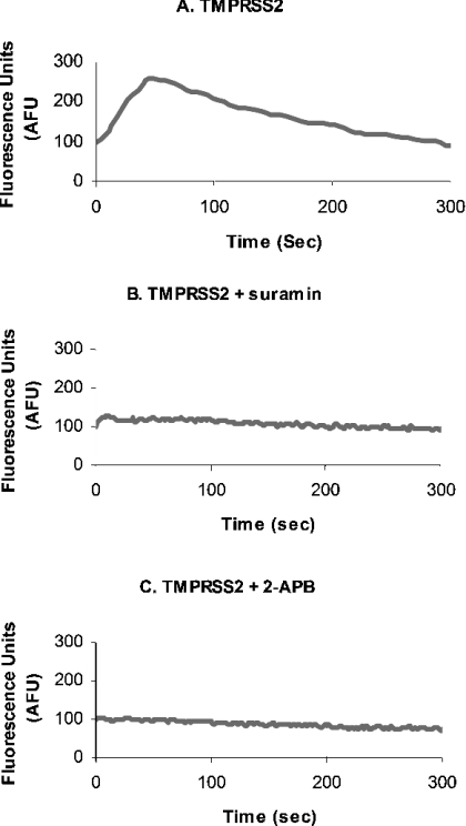
TMPRSS2 is a type II transmembrane-bound serine protease that has gained interest owing to its highly localized expression in the prostate and its overexpression in neoplastic prostate epithelium. Once activated, the serine protease domain of TMPRSS2 is released from the cell surface into the extracellular space. PAR (protease-activated receptor)-2 belongs to a family of G-protein-coupled receptors (PAR-1-4) that are activated by specific serine proteases, which are expressed in many normal and malignant cell types. Previous in vitro studies on prostate cancer cells suggest a role for PAR-2 in prostate cancer metastasis. A polyclonal anti-human TMPRSS2 antibody was generated against the TMPRSS2 serine protease domain. The antibody showed specific reactivity with recombinant expressed TMPRSS2, and so was used to extract and purify the cleaved active TMPRSS2 protease from prostate cancer cells. Reverse transcriptase PCR and Western blot analysis were used to show the expression of both TMPRSS2 and PAR-2 in the androgen-dependent LNCaP prostate cancer cell line. Treatment of LNCaP cells with the cellular immunopurified TMPRSS2 protease induced a transient increase in intracellular calcium, which is indicative of G-protein-coupled-receptor activation. This calcium mobilization was inhibited by cellular pre-treatment with a specific PAR-2 antagonist, but not with a PAR-1 antagonist; inhibition of the protease activity also failed to mobilize calcium, suggesting that TMPRSS2 is capable of cleaving and thereby activating the PAR-2 receptor. The calcium mobilization was also inhibited by cellular pre-treatment with suramin or 2-APB (2-aminoethoxydiphenyl borate), indicating that a G-protein pathway is involved and that subsequent calcium release is mainly from intracellular stores. The present study describes how TMPRSS2 may contribute to prostate tumour metastasis via the activation of PAR-2.


Figures

Figure 1
Figure 1. Detection of TMPRSS2 by RT-PCR, SDS/PAGE and Western blotting
(A) RT-PCR analysis of TMPRSS2 mRNA distribution in PC-3, DU 145 and LNCaP human prostate cell lines and RWPE-1 human transformed prostate cell line. Predicted size of reaction product for TMPRSS2 is 3.8 kb. Upper panel: Lane 1, 50 bp cDNA ladder; lane 2, PC-3 RT-PCR product (380 bp); lane 3, DU145 RT-PCR product (none detected); lane 4, LNCaP RT-PCR product (380 bp); lane 5, RWPE-1 RT-PCR product (380 bp). Lower panel: lane 2, actin control PC-3 RT-PCR product (500 bp); lane 3, actin control DU145 RT-PCR product (500 bp); lane 4, actin control LNCaP RT-PCR product (500 bp); lane 5, actin control RWPE-1 RT-PCR product (500 bp). (B) Immunoblot analysis of recombinant TMPRSS2 extracellular domain containing a T7 tag. Lane 1, See Blue Plus 2 Pre-stained Standard molecular mass markers. Samples (1 μg) of recombinant expressed TMPRSS2 were analysed by SDS/PAGE and protein stain (lane 2) or Western blot (lanes 3–5). Lane 3 was developed with polyclonal anti-TMPRSS2 antibody (1/500), lane 4 was developed with anti-T7 tag monoclonal antibody (1/10000). A major band was detected at approx. 50 kDa in both lanes. Lane 5 was developed with polyclonal anti-TMPRSS2 antibody (1/500) pre-absorbed with antigen before use. (C) SDS/PAGE analysis of cellular immunopurified TMPRSS2. Lane 1. molecular mass standards. TMPRSS2 (3.75 μg of total protein) was analysed by SDS/PAGE and protein stain (lane 2), and Western blot (lanes 3 and 4). Bands were developed using polyclonal anti-TMPRSS2 antibody (1/500). Lane 4 shows polyclonal anti-TMPRSS2 antibody pre-absorbed with TMPRSS2. Full-length TMPRSS2 is shown as a major band at 70 kDa, and the soluble extracellular protease domain at 32 kDa.
Figure 2
Figure 2. Cleavage of the substrate Cbz-Gly-Gly-Arg-AMC
Cleavage of the substrate Cbz-Gly-Gly-Arg-AMC (100 μM) by 1.25 μg of active cellular immunopurified TMPRSS2 (×), 0.5 ng of trypsin (▲), 1.25 μg of active TMPRSS2 in the presence of 10 μM Cbz-Lys(OPh)2 (■) or 1.25 μg of active TMPRSS2 in the presence of 200 μM PAR-2 antagonist (Phe-Ser-Leu-Leu-Arg-Tyr-NH2) (*).
Figure 3
Figure 3. Intracellular calcium mobilization via activation of PAR-2 on LNCaP cells
Cells were loaded with Fluo 4 and protease-triggered calcium release was assessed. Cells were treated with 100 ng of trypsin (A), 1.25 μg of soluble TMPRSS2 (B) (the inset shows a sample of immunopurified TMPRSS2 used for activation of PAR-2 analysed by SDS/PAGE from Figure 1C, with molecular mass sizes indicated in kDa), TMPRSS2 in the presence of PAR-2 antagonist (200 μM) (C) and PAR-1 antagonist (10 μM) (D), 100 ng of trypsin in the presence of PAR-2 antagonist (200 μM) (E), TMPRSS2 addition to cells 180 s after a PAR-2-desensitizing treatment of 50 μM SLIGKV (F), and TMPRSS2 pre-incubated with the irreversible serine protease inhibitor Cbz-Lys(OPh)2 (10 μM) (G). Treatments were added at zero-time, except in (F) when they were added at zero-time and at 180 s. Fluorescence was measured at 516 nm (with excitation at 494 nm).
Figure 4
Figure 4. Intracellular calcium mobilization via activation of PAR-2 on LNCaP cells
LNCaP cells were treated with TMPRSS2 (A) as before, and also pre-treated with 5 mM suramin (B) or 100 μM 2-APB before the addition of TMPRSS2 (C).

References

    1. Paoloni-Giacobino A., Chem H., Peitsch M. C., Rossier C., Antonarakis S. E. Cloning of the TMPRSS2 gene, which encodes a novel serine protease with transmembrane, LDLRA, and SRCR domains and maps to 21q22.3. Genomics. 1997;44:309–320. - PubMed
    1. Del Rosso M., Fibbi G., Pucci M., D'Alessio S., Del Rosso A., Magnelli L., Chiarugi V. Multiple pathways of cell invasion are regulated by multiple families of serine proteases. Clin. Exp. Metastasis. 2002;19:193–207. - PubMed
    1. Cocks T. M., Moffatt J. D. Protease-activated receptors: sentries for inflammation? Trends Pharmacol. Sci. 2000;21:103–108. - PubMed
    1. Darmoul D., Gratio V., Devaud H., Lehy T., Laburthe M. Aberrant expression and activation of the thrombin receptor protease-activated receptor-1 induces cell proliferation and motility in human colon cancer cells. Am. J. Pathol. 2003;162:1503–1513. - PMC - PubMed
    1. Hooper J. D., Clements J. A., Quigley J. P., Antalis T. M. Type II transmembrane serine proteases: insights into an emerging class of cell surface proteolytic enzymes. J. Biol. Chem. 2001;276:857–860. - PubMed

Publication types

MeSH terms