. 2005 Jan 1;14(1):103-11.
doi: 10.1093/hmg/ddi010. Epub 2004 Nov 10.

Digenic inheritance of deafness caused by mutations in genes encoding cadherin 23 and protocadherin 15 in mice and humans

Affiliations

Digenic inheritance of deafness caused by mutations in genes encoding cadherin 23 and protocadherin 15 in mice and humans

Qing Yin Zheng et al. Hum Mol Genet. .

Abstract

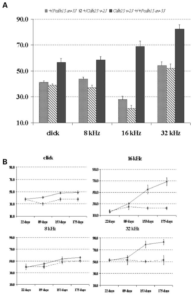
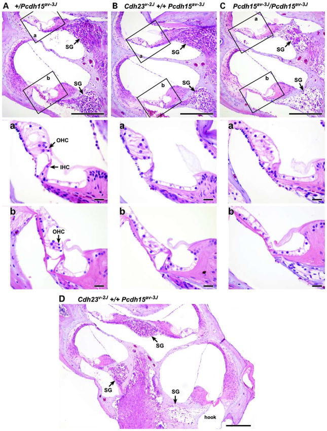
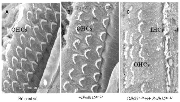
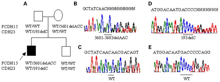
Mutations in genes coding for cadherin 23 and protocadherin 15 cause deafness in both mice and humans. Here, we provide evidence that mutations at these two cadherin loci can interact to cause hearing loss in digenic heterozygotes of both species. Using a classical genetic approach, we generated mice that were heterozygous for both Cdh23 and Pcdh15 mutations on a uniform C57BL/6J background. Significant levels of hearing loss were detected in these mice when compared to age-matched single heterozygous animals or normal controls. Cytoarchitectural defects in the cochlea of digenic heterozygotes, including degeneration of the stereocilia and a base-apex loss of hair cells and spiral ganglion cells, were consistent with the observed age-related hearing loss of these mice beginning with the high frequencies. In humans, we also have obtained evidence for a digenic inheritance of a USH1 phenotype in three unrelated families with mutations in CDH23 and PCDH15. Altogether, our data indicate that CDH23 and PCDH15 play an essential long-term role in maintaining the normal organization of the stereocilia bundle.


Figures

Figure 1
Figure 1
Elevated ABR thresholds and progression of hearing loss in double heterozygous mice. Average ABR thresholds for broadband clicks and 8, 16 and 32 kHz pure-tone stimuli are shown for 12 +/Pcdh15av-3J single heterozygotes (cross-hatched bars), five +/Cdh23v-2J single heterozygotes (diagonally marked bars) and 33 Cdh23v-2J +/+ Pcdh15av-3J double heterozygous mice (black bars) tested at 6–7 months of age. ABR threshold values below 55 (click), 50 (8 kHz), 35 (16 kHz) and 60 (32 kHz) dB SPL are considered within the normal range for mice (15). The maximum amplitude presented for each test stimulus was 100 dB. Standard errors are indicated for each threshold mean (A). For each test stimulus, the average ABR thresholds of two +/Pcdh15av-3J single heterozygous controls (squares connected by dashed line) and seven Cdh23v-2J +/+ Pcdh15av-3J double heterozygotes (diamonds connected by solid line) are plotted for successive tests performed at 22, 89, 153 and 175 days of age. Standard errors are indicated for each threshold mean (B).
Figure 2
Figure 2
Loss of hair cells and SGCs in cochlea of mutant mice. Cross-sections through three apico-basal regions of cochlea 6 months of age are shown: mid-apex (apical turn of mid-modilar section, but below the apex), mid-base (basal turn of mid-modiolar section) and the extreme base. Cochlea of a +/Pcdh15av-3J single heterozygote control (A), a Cdh23v-2J +/+ Pcdh15av-3J double heterozygote (B) and a Pcdh15av-3J/Pcdh15av-3J homozygote (C). The middle and the lower panels show magnified images corresponding to the boxed areas a (mid-apex region) and areas b (mid-base region) in the upper panels, respectively. Note the loss of hair cells (lower panels) and loss of neurons (upper panels) from both the double heterozygote and the single homozygote cochlea. SGC loss is more pronounced in the hook (extreme base) of Cdh23v-2J +/+ Pcdh15av-3J double heterozygote (D) at different magnification. SG, spiral ganglion; IHC, inner hair cell; OHC, outer hair cell. Scale bars, 200 μm for upper panels and 20 μm for middle and lower panels.
Figure 3
Figure 3
Stereocilia defect and loss of sensory hair cells of the Cdh23v-2J +/+ Pcdh15av-3J double heterozygote. Scanning electron micrographs of hair cell stereocilia in cochlea of 7-month-old mice. Stereocilia on the three rows of OHCs in a cochlea of a wild-type littermate B6 +/+ mouse (A) and of a +/Pcdh15av-3J single heterozygous mouse (B), with a normal, highly organized pattern, when compared with stereocilia of a Cdh23v-2J +/+ Pcdh15av-3J double heterozygous mutant mouse shown in (C). The single row of inner hair cells is seen in (C). Hair cells loss in the double heterozygous mutants underlies the disrupted appearance of the hair cell pattern. OHC, outer hair cell; IHC, inner hair cell. Scale bar, 5 μm.
Figure 4
Figure 4
ERG waveforms from a 6-month-old Cdh23v-2J +/+ Pcdh15av-3J double heterozygous mutant (A and B) and from an aged-matched +/Pcdh15av-3J single heterozygote control (C and D). The amplitude and implicit time of both the rod- and cone-mediated ERG are normal. Rod-dominated responses were recorded to short-wavelength (λmax = 470 nm; Wratten 47A filter) flashes of light over a 4.0 log unit range of intensities (0.3 log unit steps) up to the maximum allowable by the photic stimulator. Cone-dominated responses were obtained with white flashes (0.3 steps) on the rod-saturating background after 10 min of exposure to the background light to allow complete light adaptation. Signals were sample every 0.8 ms over a response window of 200 ms. For each stimulus condition, responses were computer-averaged with up to 50 records averaged for the weakest signals.
Figure 5
Figure 5
Evidence for digenic inheritance of USH1 involving CDH23 and PCDH15. Pedigree and segregation of the mutations in CDH23 and PCDH15. The deaf proband is indicated by an arrow. CDH23/PCDH15 genotypes are given below the respective pedigree symbol (A). Direct sequence analysis showing the 5601delACC mutation (B) and wild-type (WT) allele (C) of PCDH15. Direct sequence analysis showing the 193delC mutation (D) and wild-type (WT) allele (E) of CDH23.

References

    1. Bolz H, von Brederlow B, Ramirez A, Bryda EC, Kutsche K, Nothwang HG, Seeliger M, del CSCM, Vila MC, Molina OP, et al. Mutation of CDH23, encoding a new member of the cadherin gene family, causes Usher syndrome typ.1D. Nat Genet. 2001;27:108–112. - PubMed
    1. Bork JM, Peters LM, Riazuddin S, Bernstein SL, Ahmed ZM, Ness SL, Polomeno R, Ramesh A, Schloss M, Srisailpathy CR, et al. Usher syndrome 1D and nonsyndromic autosomal recessive deafness DFNB12 are caused by allelic mutations of the novel cadherin-like gene CDH23. Am J Hum Genet. 2001;68:26–37. - PMC - PubMed
    1. Ahmed ZM, Riazuddin S, Bernstein SL, Ahmed Z, Khan S, Griffith AJ, Morell RJ, Friedman TB, Wilcox ER. Mutations of the protocadherin gene PCDH15 cause Usher syndrome typ.1F. Am J Hum Genet. 2001;69:25–34. - PMC - PubMed
    1. Alagramam KN, Yuan H, Kuehn MH, Murcia CL, Wayne S, Srisailpathy CR, Lowry RB, Knaus R, Van Laer L, Bernier FP, et al. Mutations in the novel protocadherin PCDH15 cause Usher syndrome type 1F. Hum Mol Genet. 2001;10:1709–1718. - PubMed
    1. Ahmed ZM, Riazuddin S, Ahmad J, Bernstein SL, Guo Y, Sabar MF, Sieving P, Riazuddin S, Griffith AJ, Friedman TB, et al. PCDH15 is expressed in the neurosensory epithelium of the eye and ear and mutant alleles are responsible for both USH1F and DFNB23. Hum Mol Genet. 2003;12:3215–3223. - PubMed

Publication types

Associated data