. 2005 Jan;16(1):178-92.
doi: 10.1091/mbc.e04-03-0258. Epub 2004 Nov 10.

Ypt31/32 GTPases and their novel F-box effector protein Rcy1 regulate protein recycling

Affiliations

Ypt31/32 GTPases and their novel F-box effector protein Rcy1 regulate protein recycling

Shu Hui Chen et al. Mol Biol Cell. 2005 Jan.

Abstract

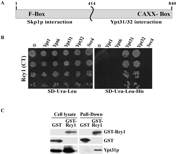
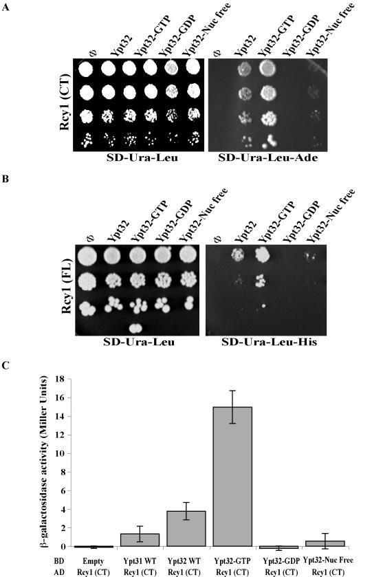
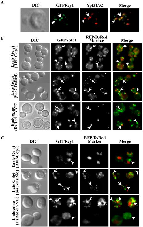
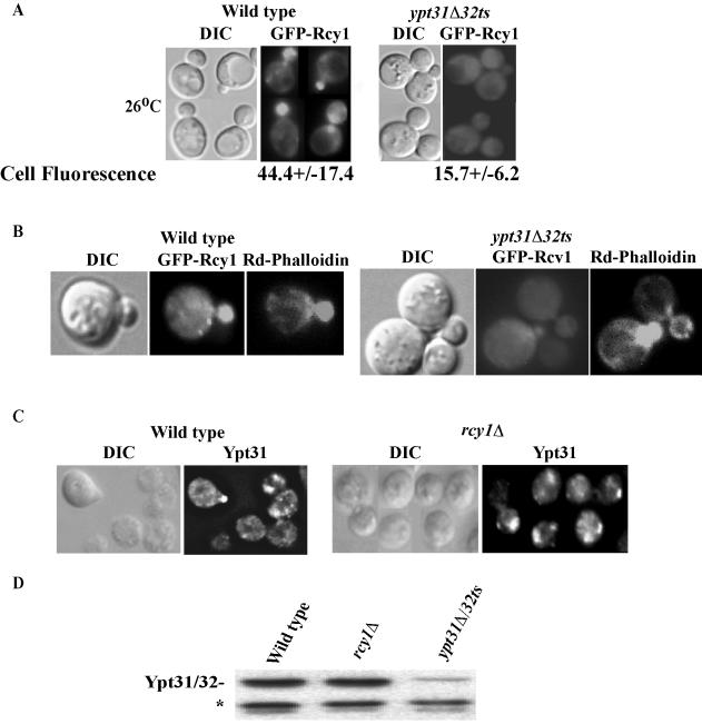
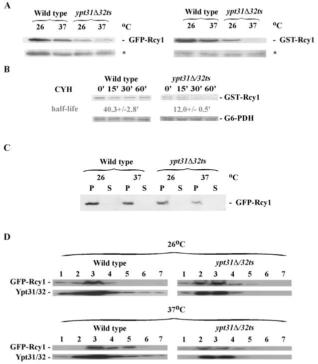
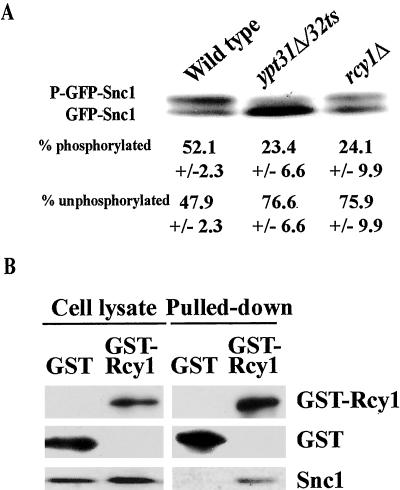
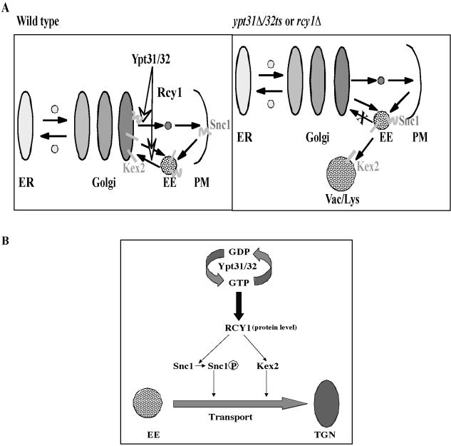
Ypt/Rab GTPases control various aspects of vesicle formation and targeting via their diverse effectors. We report a new role for these GTPases in protein recycling through a novel effector. The F-box protein Rcy1, which mediates plasma membrane recycling, is identified here as a downstream effector of the Ypt31/32 GTPase pair because it binds active GTP-bound Ypt31/32 and colocalizes with these GTPases on late Golgi and endosomes. Furthermore, Ypt31/32 regulates the polarized localization and half-life of Rcy1. This suggests that Ypt/Rabs can regulate the protein level of their effectors, in addition to the established ways by which they control their effectors. We show that like Rcy1, Ypt31/32 regulate the coupled phosphorylation and recycling of the plasma membrane v-SNARE Snc1. Moreover, Ypt31/32 and Rcy1 regulate the recycling of the furin-homolog Kex2 to the Golgi. Therefore, Ypt31/32 and Rcy1 mediate endosome-to-Golgi transport, because this is the only step shared by Snc1 and Kex2. Finally, we show that Rcy1 physically interacts with Snc1. Based on this result and because F-box proteins serve as adaptors between specific substrates and ubiquitin ligases, we propose that Ypt31/32 GTPases regulate the function of Rcy1 in the phosphorylation and/or ubiquitination of proteins that recycle through the Golgi.


Figures

Figure 1.
Figure 1.
The C-terminal domain of Rcy1 specifically interacts with Ypt31/32 GTPases. (A) Schematic diagram of RCY1 domains. The N-terminal domain contains an F-box and interacts with Skp1 (Galan et al., 2001), whereas the C-terminal domain contains a CAAX box that has been implicated in membrane localization (Galan et al., 2001) and interacts with Ypt31/32 (shown here). (B) The C-terminal domain of Rcy1p (Rcy1-CT) interacts specifically with Ypt31 and Ypt32 in the yeast two-hybrid mating assay. Yeast MATa cells expressing Rcy1 from a GAL4-AD (LEU2) vector were mated with MATα cells expressing the different exocytic Ypts from GAL4-BD (URA3) plasmids: Ypt1, Ypt6, Ypt31, Ypt32, and Sec4. The SD-Ura-Leu medium (left) supports growth of all diploid cells, whereas growth on the SD-Ura-Leu-His medium (right) requires two-hybrid interaction. (C) Rcy1 coprecipitates with Ypt31/32 from yeast cell lysates. Lysates from yeast cells expressing GST (NSY794) or GST-Rcy1 (NSY624) under the GAL1 promoter were prepared. Glutathione beads were used to pull down GST or GST-Rcy1. Cell lysates (two left lanes, 25% of the cell lysate used for pull-down) and GST-pull down fraction (two right lanes) were analyzed by Western blot. GST proteins were detected using an affinity-purified polyclonal anti-GST antibody, and Ypt31/32 proteins were detected using a polyclonal anti-Ypt31/32 antibody. Results shown in this figure are representative of at least two experiments.
Figure 2.
Figure 2.
Rcy1p interacts specifically with the GTP-bound form of Ypt31/32 GTPases. The C-terminal domain of Rcy1 (Rcy1-CT) (A) full-length Rcy1 (Rcy1-FL) (B) specifically interact with activated Ypt32 in the yeast two-hybrid mating plate assay. Yeast MATa cells expressing Rcy1 from the GAL4-AD (LEU2) vector were mated with MATα cells expressing the various nucleotide-bound forms of Ypt32 from GAL4-BD (URA3) plasmids: wild type, Q72L (GTP), S27D (GDP), and N126I (nucleotide free). Growth of the diploid cells on SD-Ura-Leu is shown on the left, whereas interaction is shown on the right as growth on SD-Ura-Leu-Ade in A, and SD-Ura-Leu-His in B (results shown in A and B are representative of at least two experiments). (C) The C-terminal domain of Rcy1 (Rcy1-CT) specifically interacts with Ypt32-GTP in the yeast two-hybrid quantitative assay. Diploid cells expressing GAL4-AD-Rcy1-CT and GAL4-BD-Ypts, Ypt31, and different nucleotide-bound forms of Ypt32 from A were tested for the strength of interactions by using a β-galactosidase liquid assay. The results represent two independent experiments; the error bars represent SD.
Figure 3.
Figure 3.
Ypt31/32 colocalize with Rcy1, and they both reside on late Golgi and endosomes. (A) GFP-Rcy1 and Ypt31/32 colocalize. Wild-type cells (NSY125) expressing GFP-Rcy1 were grown at 26°C and synchronized using α-factor (see Materials and Methods). The cells were fixed and Ypt31/32 localization was viewed by indirect immunofluorescence microscopy by using affinity-purified anti-Ypt31/32 antibodies (red). GFP-Rcy1 was viewed using an FITC filter (green). (B) Ypt31 colocalizes with Golgi and endosomal markers. Cells expressing EGFP-Ypt31 and RFP-Cop1, Sec7-DsRED, or DsRED-FYVE were viewed by direct fluorescence microscopy. EGFP-Ypt31was viewed using an FITC filter (green) and RFP- or DsRed-tagged markers were viewed using a Texas Red filter (red). (C) Rcy1 colocalizes with late Golgi and endosomal markers. Cells expressing GFP-Rcy1 and RFP-Cop1, Sec7-DsRed, or DsRED-FYVE were viewed by direct fluorescence microscopy. GFP-Rcy1 was viewed using an FITC filter (green) and RFP- or DsRed-tagged markers were viewed using a Texas Red filter (red). The images in A, B, and C were deconvolved using a Zeiss Axioscope microscope. The merged image is shown on the right, and the contour of the cells is shown in the differential interference contrast (DIC) image. Arrows point to regions of colocalization, whereas arrowheads point to regions of Ypt31/32 or Rcy1 (green) that do not colocalize with the compartmental markers (red).
Figure 4.
Figure 4.
Ypt31/32 affect the localization and the protein level of Rcy1 but not vice versa. (A) Ypt31/32 affect the localization of Rcy1. GFP-Rcy1 was expressed in wild-type (NSY125) and ypt31Δ/32ts mutant (NSY348) cells growing at 26°C. Cells were synchronized by incubation with α-factor (see Materials and Methods for details). GFP-Rcy1 protein localization was then examined by direct fluorescence microscopy. Bottom, cell fluorescence was quantified in Photoshop 7.0. Eleven random cells were quantified for each strain; ± represents the SEM. (B) Actin polarization is normal in ypt31Δ/32ts cells. Cells expressing GFP-Rcy1 were grown at 26°C, fixed, and stained with rhodamine-phalloidin, and then cells were examined by direct fluorescence microscopy. GFP-Rcy1 was viewed using an FITC filter and actin staining was viewed using a Texas Red filter. (C)Ypt31 localization is not disrupted in rcy1Δ mutants. Exponentially growing wild-type (NSY125) and rcy1Δ (NSY657) cells were fixed and Ypt31/32 localization was determined using affinity-purified anti-Ypt31p antibody and indirect immunofluorescence microscopy. In panels A–C, the contour of the cells is shown in the DIC image. (D) The level of Ypt31/32 is not affected by Rcy1. Protein level of Ypt31/32, in wild-type (NSY125), rcy1Δ (NSY657), and ypt31Δ/32ts (NSY348) cells was detected by Western blot analysis by using affinity-purified anti-Ypt31/32 antibody. Equal loading was confirmed by Ponceau S staining and a cross-reacting band that is present in ypt31Δ cells (*).
Figure 5.
Figure 5.
Ypt31/32 control the protein stability of Rcy1. (A) The level of Rcy1 protein is reduced in ypt31Δ32ts mutant cells. Wild-type (NSY125) and ypt31Δ/32ts mutant (NSY348) cells expressing GFP-Rcy1 (left) or GST-Rcy1 (right) were grown at 26°C and GFP- or GST-Rcy1 expression was induced for 5 h. After 4 h, half of each culture was shifted to 37°C for 1 h. Cell lysates were prepared and tested by Western blot analysis by using anti-GFP or anti-GST antibodies. Total cell lysate (100 μg, as determined by Bio-Rad assay) was loaded on each lane. Equal loading was confirmed by Ponceau S staining and a cross-reacting band that is present in cells containing the empty vector (*). (B) GST-Rcy1 is unstable in ypt31Δ/32ts mutant cells. Wild-type and mutant cells expressing GST-Rcy1 were grown as in A, except that cultures were shifted to 37°C 3 h after the induction of GST-Rcy1 expression. Cells were pulsed at 37°C with 35S-Translabel for 30 min, chased for the indicated times, and samples were analyzed as described in Materials and Methods. The half-life of GST-Rcy1 is indicated; ± represents the SEM. (C) GFP-Rcy1 protein is found in the P13 pellet fraction of wild-type and ypt31Δ/32ts mutant cells. Cell lysates were centrifuged at 13,000 × g for 30 min to generate pellet (P) and supernatant (S) fractions that were tested by Western blot analysis by using anti-GFP antibody. The results shown in this figure are representative of three experiments. (D) Rcy1 and Ypt31/32 in the P13 fraction are associated with membranes. P13 fractions from part C were analyzed on iodixanol density gradients; gradient fractions were collected and tested by Western blot analysis. Membranes fractionate to the top of the gradient. The results shown in this figure are representative of at least two independent experiments.
Figure 6.
Figure 6.
Ypt31/32 and Rcy1 play a role in the recycling of the Kex2 and Snc1 proteins. (A) Ypt31/32 and Rcy1 affect Kex2 stability. Wild-type (NSY125), ypt31Δ/32ts (NSY348), ypt31Δ/32ts pep4Δ (NSY355), rcy1Δ (NSY818), and rcy1Δ pep4Δ (NSY819) cells expressing HA-tagged Kex2 protein were grown entirely at 26°C or shifted to 37°C for 90 min before harvest as indicated. Cell lysates were prepared and Kex2 protein level was measured by Western blot analysis by using anti-HA antibodies. Total protein determination and Emp47 were used as loading and blotting controls. Typical results representing five experiments are shown. (B) Microscopic assay showing that Ypt31/32 GTPases are important for the recycling of Kex2 to the Golgi. Wild-type (NSY970), pep4Δ (NSY971), ypt31Δ/32ts (NSY972), and ypt31Δ/32ts pep4Δ (NSY973) cells expressing endogenous Kex2 tagged with YFP on the chromosome were grown at 26°C. FM4-64 was internalized for 30 min to mark the vacuolar membrane. Intracellular localization was determined by direct fluorescence using an FITC filter for Kex2-YFP and Texas Red filter for FM4-64. Kex2-YFP in the Golgi is seen as green dots outside the vacuolar ring (arrows) in wild-type but not mutant cells. (C) Ypt31/32 and Rcy1 are involved in Snc1 recycling to the plasma membrane. Wild-type (NSY729), ypt31Δ/32ts (NSY733), and rcy1Δ (NSY737) cells expressing GFP-tagged Snc1 protein were grown at 26°C. GFP-Snc1 intracellular localization was determined by direct fluorescence by using an FITC filter. GFP-Snc1-pem, which cannot be internalized, is shown at the bottom. This form of Snc1 localizes to the PM in wild-type and mutant cells. (D) Snc1 localizes to early endosomes in ypt31Δ/32ts and rcy1Δ mutant cells. Cells were grown and treated as in C, except that early endosomes were marked by FM4-64 internalized for 5 min (some FM4-64 is still on the plasma membrane after such a short pulse). In C and D, arrows indicate PM staining, and arrowheads point to internalized GFP-Snc1; the contour of the cells is shown in the DIC image.
Figure 7.
Figure 7.
Rcy1 interacts with Snc1 and together with Ypt31/32 affects the level of Snc1 phosphorylation. (A) Ypt31/32 and Rcy1 affect the phosphorylation state of Snc1. Wild-type and mutant cells expressing GFP-Snc1 were grown at 26°C. Cell lysates were prepared and assayed by Western blot analysis by using anti-Snc1 antibodies. The higher molecular weight band is the phosphorylated form of Snc1, whereas the lower molecular weight band is the unphosphorylated form (Galan et al., 2001). The bands were quantified using an Alpha Imager, and the percentage of phosphorylated Snc1 averaged from three independent experiments is indicated; ± represents the SEM. (B) Rcy1 interacts with Snc1. Cell lysates were prepared from wild-type cells expressing GST (NSY784) or GST-Rcy1 (NSY624) as described in Materials and Methods. Cell lysates (two left lanes, 25% of the cell lysate used for pull-down) and GST-pull down fraction (two right lanes) were assayed by Western blot analysis. GST proteins were detected using a polyclonal anti-GST antibody, and Snc1 protein was detected using a polyclonal anti-Snc1 antibody. The results shown are representative of two experiments. Snc1 occurs as two bands in A and one band in B because the separation of the two bands requires electrophoresis in a high percent gel (15%) for a long period (overnight at 4°C), which was not done in B.
Figure 8.
Figure 8.
Proposed role of Ypt31/32 GTPases in the regulation of protein recycling through the Golgi. (A) Model of the transport step regulated by Ypt31/32 and Rcy1. We suggest that Ypt31/32 and its effector Rcy1 regulate endosome-to-Golgi recycling. In wild-type cells (left), Kex2 cycles between the trans-Golgi and early endosomes, whereas the v-SNARE Snc1 is endocytosed into early endosomes and is transported back to the PM via the trans-Golgi. In ypt31Δ/32ts and rcy1Δ mutant cells (right), endosome-to-Golgi transport is blocked. In these cells, Kex2 is transported to the vacuole where it is degraded, whereas most of the Snc1 accumulates in early endosomes. (B) Model of the mechanism by which the Ypt31/32 GTPases and their effector Rcy1 regulate the sorting and marking of proteins that are destined to be transported through the Golgi. In this model, Ypt31/32 GTPases, in their active form, bind their effector Rcy1 and modulate its protein level. They may also regulate its membrane recruitment and/or activity. The Rcy1 F-box protein, in turn, binds the recycling proteins Snc1 and Kex2 and mediates their marking for recycling by phosphorylation. This phosphorylation might be followed by ubiquitination via a non-SCF complex.

References

    1. Benli, M., Doring, F., Robinson, D. G., Yang, X., and Gallwitz, D. (1996). Two GTPase isoforms, Ypt31p and Ypt32p, are essential for Golgi function in yeast. EMBO J. 15, 6460-6475. - PMC - PubMed
    1. Blackwell, E., Halatek, I. M., Kim, H. J., Ellicott, A. T., Obukhov, A. A., and Stone, D. E. (2003). Effect of the pheromone-responsive G(alpha) and phosphatase proteins of Saccharomyces cerevisiae on the subcellular localization of the Fus3 mitogen-activated protein kinase. Mol. Cell. Biol. 23, 1135-1150. - PMC - PubMed
    1. Brickner, J. H., and Fuller, R. S. (1997). SOI1 encodes a novel, conserved protein that promotes TGN-endosomal cycling of Kex2p and other membrane proteins by modulating the function of two TGN localization signals. J. Cell Biol. 139, 23-36. - PMC - PubMed
    1. Byers, B. (1981). Cytology of the yeast life cycle. In: The Molecular Biology of the Yeast Saccharomyces, ed. E.J.J.N. Strathern, and J. Broach, Cold Spring Harbor, NY: Cold Spring Harbor Laboratory Press, 59-96.
    1. Carminati, J. L., and Stearns, T. (1997). Microtubules orient the mitotic spindle in yeast through dynein-dependent interactions with the cell cortex. J. Cell Biol. 138, 629-641. - PMC - PubMed

Publication types

MeSH terms