. 2004 Nov 23;101(47):16659-64.
doi: 10.1073/pnas.0407643101. Epub 2004 Nov 10.

Histone deacetylase inhibition-mediated neuronal differentiation of multipotent adult neural progenitor cells

Affiliations

Histone deacetylase inhibition-mediated neuronal differentiation of multipotent adult neural progenitor cells

Jenny Hsieh et al. Proc Natl Acad Sci U S A. .

Abstract

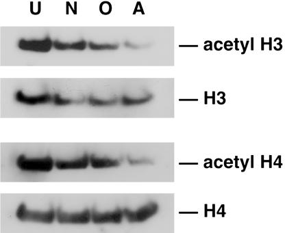
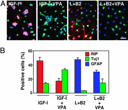
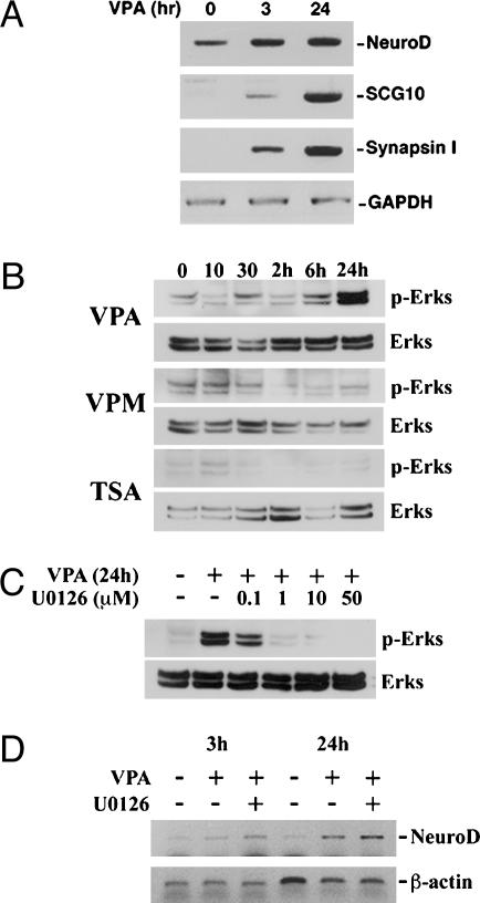
It has become apparent that chromatin modification plays a critical role in the regulation of cell-type-specific gene expression. Here, we show that an inhibitor of histone deacetylase, valproic acid (VPA), induced neuronal differentiation of adult hippocampal neural progenitors. In addition, VPA inhibited astrocyte and oligodendrocyte differentiation, even in conditions that favored lineage-specific differentiation. Among the VPA-up-regulated, neuron-specific genes, a neurogenic basic helix-loop-helix transcription factor, NeuroD, was identified. Overexpression of NeuroD resulted in the induction and suppression of neuronal and glial differentiation, respectively. These results suggest that VPA promotes neuronal fate and inhibits glial fate simultaneously through the induction of neurogenic transcription factors including NeuroD.


Figures

Fig. 1.
Fig. 1.
HDAC-inhibition-mediated neuronal differentiation. (A) Treatment of neural progenitors with 1 mM VPA for 4 days resulted in an increase in cells with MAP2ab (green) staining and neuronal morphology compared with control cultures, which lack neuronal differentiation. Similar results were observed with the addition of 100 nM TSA and 1 μM NaB for 2 days. Blue regions indicate 4′,6-diamidino-2-phenylindole-stained nuclei. (B) Staining of acetylated histone H3 (red) and MAP2ab (green) is higher in HDAC inhibitor-treated cultures. (C) Proliferation in untreated (control) and 1-day HDAC inhibitor-treated cultures as determined by BrdUrd (red) incorporation. (Scale bar, 50 μm.) (D and E) Quantifications of neuronal differentiation (D) and proliferation (E) in untreated and HDAC inhibitor-treated cultures. All data shown are from at least three experiments in parallel cultures with error bars representing standard deviations.
Fig. 2.
Fig. 2.
Changes in histone acetylation during neural progenitor lineage progression. Western blot analysis of neural progenitor extracts from undifferentiated (U) cultures grown in N2 media plus 20 ng/ml FGF-2 or differentiated with 1 μM retinoic acid plus 5 μM forskolin for 4 days (neuron, N), 500 ng/ml IGF-1 for 4 days (oligodendrocyte, O), or 50 ng/ml LIF plus 50 ng/ml BMP-2 for 6 days (astrocyte, A). Immunoblotting was performed by using antibodies against histones H3 and H4 and acetylated histones H3 and H4. Data shown are from at least three independent experiments.
Fig. 3.
Fig. 3.
VPA-mediated suppression of glial differentiation. (A) Neural progenitors induced to differentiate into Rip-positive oligodendrocytes (red) with 500 ng/ml IGF-1 or GFAP-positive astrocytes (red) with 50 ng/ml LIF (L) plus 50 ng/ml BMP-2 (B2). Treatment of cultures with 1 mM VPA for 4 days reduced the number of Rip-positive cells (red) in IGF-1-treated cultures and GFAP-positive cells (red) in LIFplusBMP-2-treated cultures, while increasing the number of Tuj1-positive cells (green) in each case. (Scale bar, 50 μm.) (B) Quantification of neurons, oligodendrocytes, and astrocytes in various differentiation conditions in the presence or absence of 1 mM VPA. All data shown are from at least three experiments in parallel cultures with error bars representing standard deviations.
Fig. 4.
Fig. 4.
VPA up-regulates neuron-specific genes, including the bHLH transcription factor NeuroD.(A) RT-PCR of NeuroD, SCG10, and Synapsin I in neural progenitors at time 0 or treated with 1 mM VPA for 3 h and 24 h. GAPDH was used as a normalization control. (B) Western blot time-course analysis of ERK activation after 1 mM VPA, 100 nM TSA, and 1 mM valpromide (VPM) treatment. (C) ERK activation with 1 mM VPA treatment with and without addition of U0126 (different doses are indicated). (D) RT-PCR analysis of VPA-mediated up-regulation of NeuroD (3 and 24 h) with and without U0126 (1 μM).
Fig. 5.
Fig. 5.
VPA decreases granule cell proliferation and increases neuronal differentiation in the dentate gyrus of adult rats. (A) Schematic of VPA and BrdUrd injection paradigm. (B) Representative images of brain sections focusing on the dentate gyrus (DG) of animals injected with saline (control) or VPA. Sections were processed for BrdUrd staining. White arrow indicates the dentate gyrus. Hi, Hilus. (Scale bar, 200 μm.) (C) The average number of BrdUrd-positive cells (in four adjacent fields) per section (control animals, n = 4; VPA-treated animals, n = 6) is plotted. The asterisk indicates that values are significantly different between control and VPA-treated animals (P < 0.001, t test). (D) The percentage of Tuj1-/BrdUrd-double positive cells (in four adjacent fields) per section (control animals, n = 4; VPA-treated animals, n = 6) is plotted. Asterisks indicate statistical significance (P < 0.05, t test). Error bars represent standard deviations.

References

    1. Georgopoulos, K. (2002) Nat. Rev. Immunol. 2, 162–174. - PubMed
    1. Grunstein, M. (1997) Nature 389, 349–352. - PubMed
    1. Kuo, M. H. & Allis, C. D. (1998) BioEssays 20, 615–626. - PubMed
    1. Struhl, K. (1998) Genes Dev. 12, 599–606. - PubMed
    1. Jenuwein, T. & Allis, C. D. (2001) Science 293, 1074–1080. - PubMed

Publication types

MeSH terms

Substances

LinkOut - more resources