. 2004 Nov-Dec;11(6):787-93.
doi: 10.1101/lm.77004. Epub 2004 Nov 10.

Central localization of plasticity involved in appetitive conditioning in Lymnaea

Affiliations

Central localization of plasticity involved in appetitive conditioning in Lymnaea

Volko A Straub et al. Learn Mem. 2004 Nov-Dec.

Abstract

Learning to associate a conditioned (CS) and unconditioned stimulus (US) results in changes in the processing of CS information. Here, we address directly the question whether chemical appetitive conditioning of Lymnaea feeding behavior involves changes in the peripheral and/or central processing of the CS by using extracellular recording techniques to monitor neuronal activity at two stages of the sensory processing pathway. Our data show that appetitive conditioning does not affect significantly the overall CS response of afferent nerves connecting chemosensory structures in the lips and tentacles to the central nervous system (CNS). In contrast, neuronal output from the cerebral ganglia, which represent the first central processing stage for chemosensory information, is enhanced significantly in response to the CS after appetitive conditioning. This demonstrates that chemical appetitive conditioning in Lymnaea affects the central, but not the peripheral processing of chemosensory information. It also identifies the cerebral ganglia of Lymnaea as an important site for neuronal plasticity and forms the basis for detailed cellular studies of neuronal plasticity.


Figures
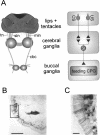
Figure 1.
Figure 1.
Organization of lip and tentacle sensory pathways to the CNS. (A) Schematic diagram of anatomical organization of lip and tentacle sensory pathways. The lips and tentacles are connected to the cerebral ganglia via the median lip nerve (mln), superior lip nerve (sln), and tentacle nerve (tn). The cerebral-buccal connective (cbc) forms the only connection between the cerebral and buccal ganglia that contain the feeding CPG. Primary chemosensory cells in the lips and tentacles extend direct projections in the lip and tentacle nerves that synapse onto sensory integrating neurons (SI) in the cerebral ganglia (for simplicity, interactions are shown as direct connections). These neurons affect the feeding CPG in the buccal ganglia via the cerebral-buccal connective. (B) Photo-micrograph of putative primary chemosensory cells labeled by backfilling the median lip nerve in a lip section. The picture shows biocytin-labeled neuronal cell bodies arranged in a subepithelial layer. Scale bar, 100 μm. (C) Enlargement of area marked by square in B. Note the fine processes originating from the labeled subepithelial cell bodies that project through the lip epithelium. Scale bar, 20 μm.
Figure 2.
Figure 2.
Sensory nerves in naive snails show similar responses to sucrose and AA application to the lips. (A) Simultaneous recordings from median lip nerve (mln), superior lip nerve (sln), and tentacle nerve (tn) showing responses of the three nerves to sucrose (0.02 M, 30 sec) applied to the lips. Note that the lip and tentacle nerves showed a considerable level of spontaneous activity that varied to some extent between individual preparations, including both tonic firing and spontaneous bursts of activity. The level of spontaneous activity did not influence the overall responses of the nerves to chemical stimulation of the lips. All nerves to the lips were cut to remove efferent influences from the CNS. (B) Equivalent record from another preparation showing responses of the same three nerves to AA (0.54 mM, 30 sec) applied to the lips. It should be noted that the two unidentified larger units that fire at an almost constant frequency throughout the record contributed only a small proportion to the overall nerve activity and that the main change in activity occurred in the small to medium sized units. (C,D) Average time course of changes in nerve firing rates in median lip nerve, superior lip nerve and tentacle nerve in response to sucrose (C, n = 7 preparations; error bars not shown for clarity) and AA (D, n = 7 preparations; error bars not shown for clarity) application.
Figure 3.
Figure 3.
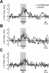
Sensory nerve responses to lip stimulation with AA are not significantly different in preparations from control and conditioned snails. (A,B,C) Average change in nerve activity in response to AA (0.54 mM, 30 sec) applied to the lips recorded in the median lip nerve (A), superior lip nerve (B), and tentacle nerve (C) of control and conditioned snails (n = 7 for each nerve; error bars not shown for clarity).
Figure 4.
Figure 4.
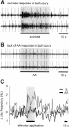
Sucrose, but not AA, application to the lips results in an increase of cerebral-buccal connective spike activity in preparations from naive snails. (A) Simultaneous recording from both cut cerebral-buccal connectives showing the response of the two connectives to sucrose (0.02 M, 30 sec) applied to the lips. (B) Record from another preparation showing the lack of a cerebral-buccal connective response to AA (0.54 mM, 30 sec) applied to the lips in a naive preparation. (C) Time course of the average changes of spike activity in the cerebral-buccal connective in response to sucrose (n = 5 preparations) and AA (n = 7 preparations) in naive snails (error bars not shown for clarity). The difference in the average frequency change during the 30 sec of stimulus application was statistically significant.
Figure 5.
Figure 5.
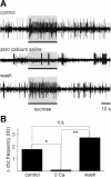
Response of cerebral-buccal connective to lip application of sucrose is blocked by zero calcium saline, suggesting that it is mediated by chemical synapses in the cerebral ganglia. (A) Sample records from a cut cerebral-buccal connective showing responses to sucrose (0.02 M, 30 sec) applied to the lips in normal saline (top trace), in nominally zero calcium saline (middle trace), and after return to normal saline (bottom trace). Note the reduced levels of spontaneous activity and the lack of an obvious response to sucrose application in zero calcium saline. (B) The summary of average changes in cerebral-buccal connective activity during the period of sucrose application shows that zero calcium saline applied to the cerebral ganglia completely abolishes the sucrose response in the cerebral-buccal connective (one-way ANOVA for correlated samples, P < 0.01 followed by post-hoc Tukey HSD tests, control versus zero calcium: P < 0.05, zero calcium versus wash: P < 0.01; n = 6 preparations).
Figure 6.
Figure 6.
Appetitive conditioning enhances cerebral-buccal connective response to AA stimulation of the lips. (A) Simultaneous recordings from the left and right cerebral-buccal connective in a preparation made from a control animal shows the lack of a response during application of AA. This is comparable to the situation in naive snails (see Fig. 4B). (B) Equivalent recordings from both cerebral-buccal connectives in a preparation made from a conditioned animal. Note the increase in overall activity in response to the application of AA (0.54 mM, 30 sec) to the lips. (C,D) Time course of average changes in total cerebral-buccal connective spike activity in response to the CS and US in control and conditioned animals. The total change during the 30-sec period of CS application in control animals was significantly different from the CS response in conditioned animals and the US responses in both control and conditioned animals (ANOVA, F(3,24) = 7.58, P < 0.01; post hoc Games/Howell tests, P < 0.05). Further statistical analysis showed that the time course of the cerebral-buccal connective frequency changes during the US and CS application in conditioned animals was not significantly different (ANOVA for repeated measures, P > 0.05 for within-subject effects [time], between-subject effects [group], and interaction effects [time × group]). An analysis of the time course of cerebral-buccal connective frequency changes during the US application in control and conditioned animals as well as in naive animals (Fig. 4C) also revealed no significant differences (ANOVA for repeated measures [Huynh-Feldt corrected], P > 0.05 for within-subject effects [time], between-subjects effects [groups], and interaction effects [time × groups]).

Similar articles

Cited by

References

    1. Alexander Jr., J., Audesirk, T.E., and Audesirk, G.J. 1984. One-trial reward learning in the snail Lymnaea stagnalis. J. Neurobiol. 15: 67-72. - PubMed
    1. Alkon, D.L., Sakakibara, M., Forman, R., Harrigan, J., Lederhendler, I., and Farley, J. 1985. Reduction of two voltage-dependent K+ currents mediates retention of a learned association. Behav. Neural. Biol. 44: 278-300. - PubMed
    1. Antonov, I., Antonova, I., Kandel, E.R., and Hawkins, R.D. 2001. The contribution of activity-dependent synaptic plasticity to classical conditioning in Aplysia. J. Neurosci. 21: 6413-6422. - PMC - PubMed
    1. Benjamin, P.R. and Elliott, C.J.H. 1989. Snail feeding oscillator: The central pattern generator and its control by modulatory interneurons. In Neural and cellular oscillators (ed. J.W. Jacklet), pp. 173-214. Dekker, New York.
    1. Benjamin, P.R., Staras, K., and Kemenes, G. 2000. A systems approach to the cellular analysis of associative learning in the pond snail Lymnaea. Learn. Mem. 7: 124-131. - PubMed

Publication types