. 2004 Nov 10;24(45):10047-56.
doi: 10.1523/JNEUROSCI.2583-04.2004.

Independent coding of movement direction and reward prediction by single pallidal neurons

Affiliations

Independent coding of movement direction and reward prediction by single pallidal neurons

David Arkadir et al. J Neurosci. .

Abstract

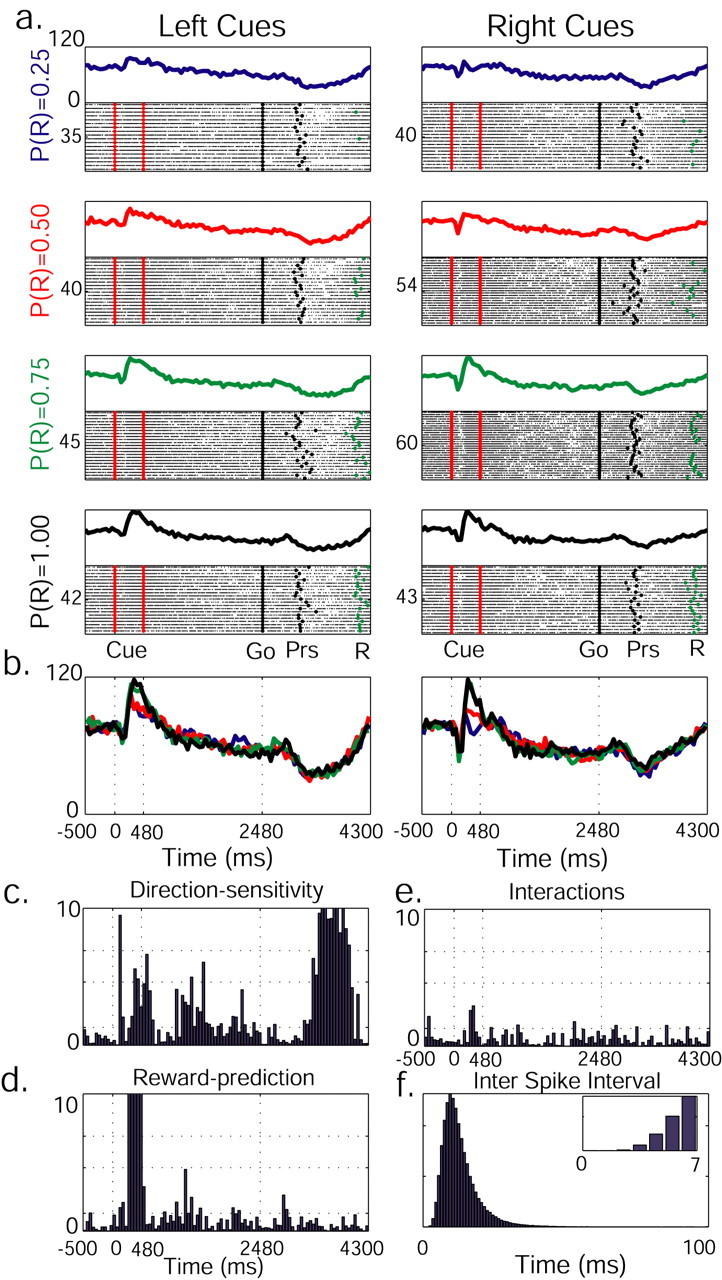
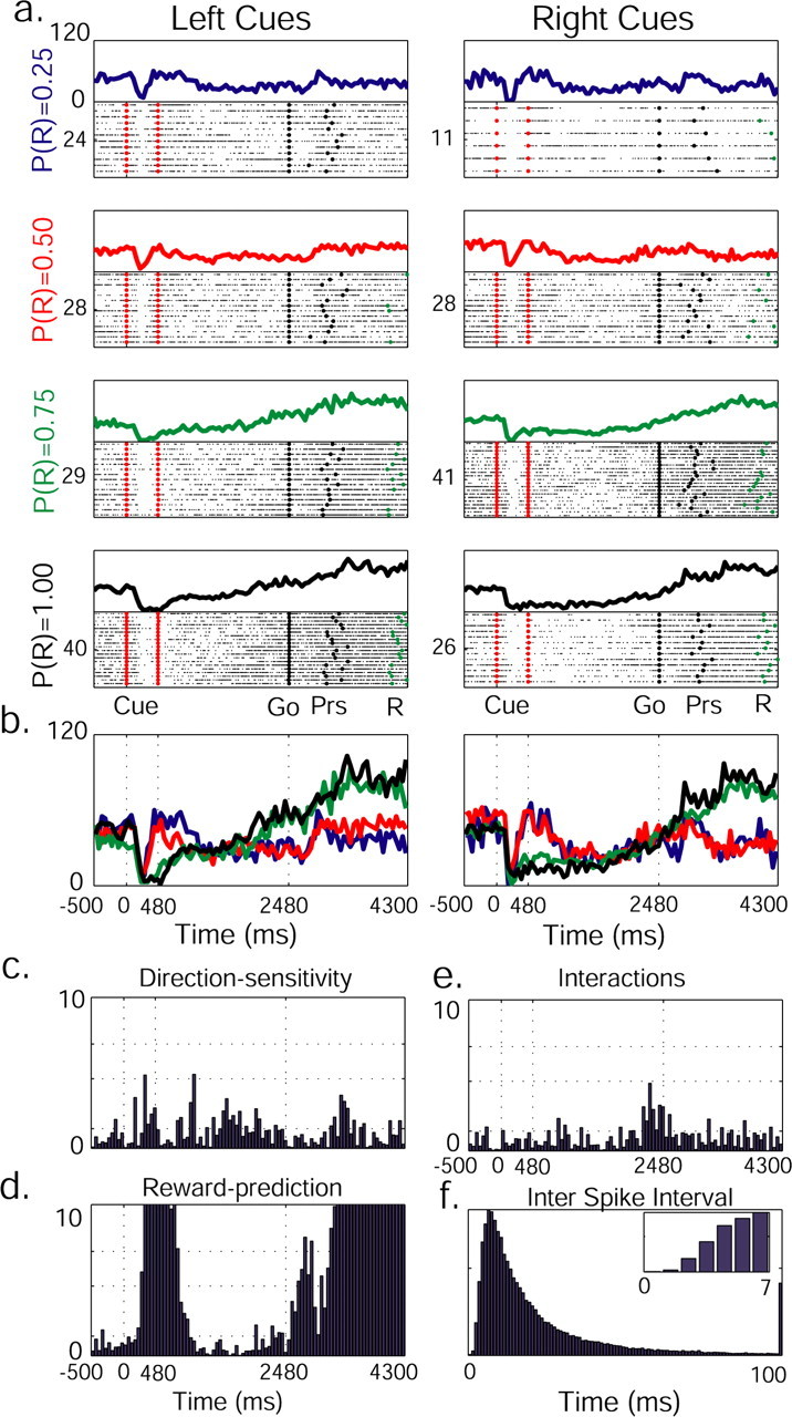
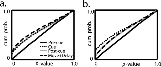
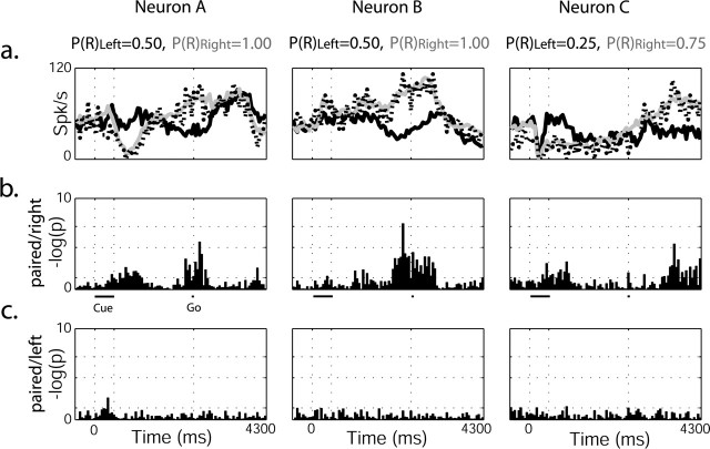
Associating action with its reward value is a basic ability needed by adaptive organisms and requires the convergence of limbic, motor, and associative information. To chart the basal ganglia (BG) involvement in this association, we recorded the activity of 61 well isolated neurons in the external segment of the globus pallidus (GPe) of two monkeys performing a probabilistic visuomotor task. Our results indicate that most (96%) neurons responded to multiple phases of the task. The activity of many (34%) pallidal neurons was modulated solely by direction of movement, and the activity of only a few (3%) pallidal neurons was modulated exclusively by reward prediction. However, the activity of a large number (41%) of single pallidal neurons was comodulated by both expected trial outcome and direction of arm movement. The information carried by the neuronal activity of single pallidal neurons dynamically changed as the trial progressed. The activity was predominantly modulated by both outcome prediction and future movement direction at the beginning of trials and became modulated mainly by movement-direction toward the end of trials. GPe neurons can either increase or decrease their discharge rate in response to predicted future reward. The effects of movement-direction and reward probability on neural activity are linearly summed and thus reflect two independent modulations of pallidal activity. We propose that GPe neurons are uniquely suited for independent processing of a multitude of parameters. This is enabled by the funnel-structure characteristic of the BG architecture, as well as by the anatomical and physiological properties of GPe neurons.


Figures

Figure 1.
Figure 1.
MRI localization of the GPe. The 2 mm MRI scans were aligned with coronal sections of an anatomical atlas of a Macaca fascicularis (Szabo and Cowan, 1984; Martin and Bowden, 2000). Each coronal plan was located relative to the anterior commissure (AC). Major structures are marked (Cd, caudate; Pt, putamen; GP, globus pallidus) with electrode (E) and recording chamber (RC). In monkey Y, the MRI scan was performed without an electrode, and recording areas were estimated only according to the borders of the recording chamber. Identification of the recording area in monkey C was also aided by an electrode that was positioned in the center of the chamber.
Figure 2.
Figure 2.
Illustration of the probabilistic visuomotor task. In response to a “go” signal, the monkey pressed the button on the same side as the remembered visual cue. In a successful trial, a reward was delivered after a delay period in accordance with the cue-encoded probability.
Figure 3.
Figure 3.
Behavior parameters of both monkeys. In a-c, data were collected from single-cue trials. The white columns represent left trials. a, Mean percentage of correct trials. b, c, Mean and SD of reaction (RT) and movement (MT) times of correct trials. d, Percentage of right button presses on double-cue trials. The meshed squares represent missing data. The behavioral and neuronal data are based on the same recording epochs.
Figure 4.
Figure 4.
The activity of a single pallidal neuron is dynamically modulated by both future reward probability and direction of movement. a, Raster plot and PSTH are presented for each reward probability and cue position. The raster plots were diluted (1:2 trials) to enhance visibility. On raster plots, significant behavioral events are marked with dots (red, cue presentation; black, “go” signal and button press; green, reward delivery). The PSTH is built of 50 msec bins. On the left of each raster plot is the number of trials. During cue display, neuronal activity increases with reward probability increment. b, PSTHs for different reward probabilities are overlaid: P(R) = 0.25 (blue), P(R) = 0.50 (red), P(R) = 0.75 (green), P(R) = 1.00 (black). c, Neuronal activity as modulated by left/right trials. A two-way ANOVA on spike counts in parallel time-lag bins was performed, and log(p) was calculated. Cue onset, offset, and “go” signal are marked with vertical dotted lines. Direction-sensitive activity during the reward-delay period might be related to the planning of the movement back to the central key in preparation for the next trial. d, Modulation of neuron activity by reward probability. e, p values reflecting interactions between the two parameters. In c-e, significance levels are marked with dotted horizontal lines (p = 0.05, p = 0.01, p = 0.001). f, Interspike interval diagram of the neuron presents a clear refractory period (see inset for higher time resolution).
Figure 5.
Figure 5.
Reward-predicting and direction-sensitive pallidal neuron. See Figure 3 for details of labeling. Note a decrease in discharge rate with the increase in reward probability (cue-display) and bipolar response (reward-delay), a combination of increased firing rates in trials in which reward was more likely, and a decrease in firing rates when reward was less likely (reward-delay).
Figure 6.
Figure 6.
Pallidal neurons convey information on probability of future reward and direction of movement. Distribution of grouped p values as calculated by a two-way ANOVA for all bins in each neuron is shown. a, Direction influence on neuronal activity is represented in the cumulative distribution of p values. Each phase on the task is marked differently. b, Influence of reward probability. The conventions are the same as in a.
Figure 7.
Figure 7.
Pallidal neurons tend to monotonically modulate their discharge rate with the change in reward probability. The relationship between firing rate and reward probability was modeled by linear regression for all bins in each reward-predicting neuron (n = 27). The cumulative distributions of linear regression p values are presented for the pre-cue period (gray) and for the cue-onset and thereafter (black).
Figure 8.
Figure 8.
Pallidal neuron ignores the irrelevant cue when two cues are presented simultaneously. a, PSTHs of three pallidal neurons (rightmost neurons also appear in Fig. 5). Black line, Single left cue; gray line, single right cue; dotted line, paired-cue trials. Reward probability values appear above. In all of the above trials, the monkey chose to press the right button, associated with the higher probability of reward. b, ANOVA on spike counts in parallel time-lag bins was performed, and log(p) was calculated and presented for the respective neurons above. Significance levels are marked with dotted horizontal lines (p = 0.05, p = 0.01, p = 0.001). There was a significant difference between the response of the neuron on left cue single trials and paired-cue trials. c, No significant difference was found for the response of the neuron on right cue single trials versus paired-cue trials.

References

    1. Albin RL, Young AB, Penney JB (1989) The functional anatomy of basal ganglia disorders. Trends Neurosci 12: 366-375. - PubMed
    1. Alexander GE, Crutcher MD (1990) Functional architecture of basal ganglia circuits: neural substrates of parallel processing. Trends Neurosci 13: 266-271. - PubMed
    1. Alexander GE, DeLong MR, Strick PL (1986) Parallel organization of functionally segregated circuits linking basal ganglia and cortex. Annu Rev Neurosci 9: 357-381. - PubMed
    1. Aosaki T, Tsubokawa H, Ishida A, Watanabe K, Graybiel AM, Kimura M (1994) Responses of tonically active neurons in the primate's striatum undergo systematic changes during behavioral sensorimotor conditioning. J Neurosci 14: 3969-3984. - PMC - PubMed
    1. Bar-Gad I, Morris G, Bergman H (2003) Information processing, dimensionality reduction and reinforcement learning in the basal ganglia. Prog Neurobiol 71: 439-473. - PubMed

Publication types

LinkOut - more resources