. 2004 Nov 10;24(45):10064-73.
doi: 10.1523/JNEUROSCI.2981-04.2004.

Axonal regeneration and lack of astrocytic gliosis in EphA4-deficient mice

Affiliations

Axonal regeneration and lack of astrocytic gliosis in EphA4-deficient mice

Yona Goldshmit et al. J Neurosci. .

Abstract

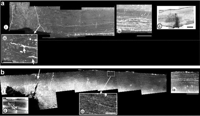
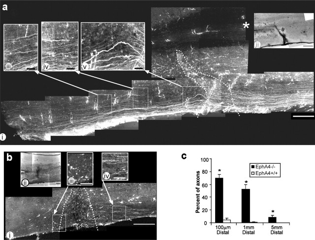
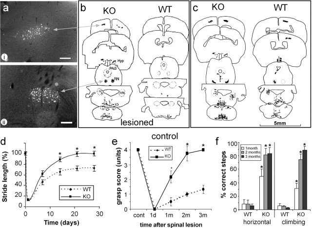
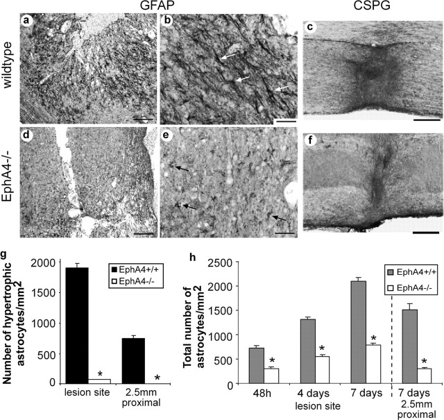
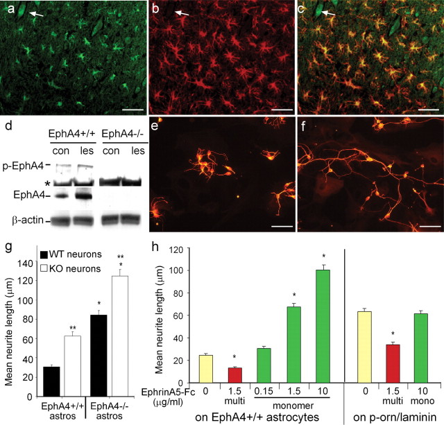
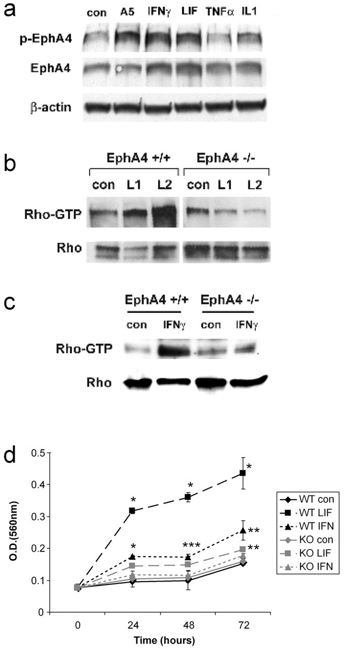
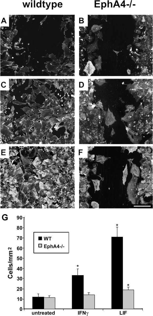
Spinal cord injury usually results in permanent paralysis because of lack of regrowth of damaged neurons. Here we demonstrate that adult mice lacking EphA4 (-/-), a molecule essential for correct guidance of spinal cord axons during development, exhibit axonal regeneration and functional recovery after spinal cord hemisection. Anterograde and retrograde tracing showed that axons from multiple pathways, including corticospinal and rubrospinal tracts, crossed the lesion site. EphA4-/- mice recovered stride length, the ability to walk on and climb a grid, and the ability to grasp with the affected hindpaw within 1-3 months of injury. EphA4 expression was upregulated on astrocytes at the lesion site in wild-type mice, whereas astrocytic gliosis and the glial scar were greatly reduced in lesioned EphA4-/- spinal cords. EphA4-/- astrocytes failed to respond to the inflammatory cytokines, interferon-gamma or leukemia inhibitory factor, in vitro. Neurons grown on wild-type astrocytes extended shorter neurites than on EphA4-/- astrocytes, but longer neurites when the astrocyte EphA4 was blocked by monomeric EphrinA5-Fc. Thus, EphA4 regulates two important features of spinal cord injury, axonal inhibition, and astrocytic gliosis.


Figures

Figure 1.
Figure 1.
At 6 d after injury, EphA4-/- axons approach but do not cross the lesion site. Anterograde tracing and confocal analysis of lesioned EphA4-/- spinal cords 6 d after hemisection (a) show large numbers of labeled axons 2.5 mm proximal to the lesion (ia) and a small number of axons with growth cones (aiii, arrows) approaching the lesion site, which is indicated by the dotted line (i) and shown more clearly in a hematoxylin and eosin (H&E)-stained section (ii). b, Wild-type spinal cord also shows very few axons approaching the lesion site. bia shows labeling 2.5 mm upstream of the lesion site. biii, An enlargement of bi shows few axons upstream of the lesion site. In both panels, rostral is to the right and caudal to the left, and the lesion site is indicated by dotted lines. Enlarged areas are indicated by boxed areas and arrows. Scale bars: i, 250 μm; ii, 200 μm; iii, 50 μm.
Figure 2.
Figure 2.
Extensive axonal regeneration in EphA4-/- mice at 6 weeks after injury. Anterograde tracing and confocal analysis of lesioned EphA4-/- spinal cords 6 weeks after hemisection showed that a large percentage of EphA4-/- axons crossed the lesion site (a, c) and extended caudally (*p < 0.001), unlike wild-type (EphA4+/+) axons that did not cross the lesion site (b, c). A montage of confocal images of EphA4-/- spinal cord (ai) showed that the regenerating axons passed through the lesion site (indicated by dotted line and by H&E-stained section in aii and extended caudally in a straight line with some “waviness” seen immediately after lesion in aiii, iv, and v). In both panels, rostral is to the right and caudal to the left, and the lesion site is indicated by dotted lines. Enlarged areas are indicated by boxed areas and arrows. ii in both cases shows an adjacent H&E-stained section demonstrating the lesion site. Scale bars: i, 250 μm; ii, 200 μm; iii—v, 50 μm. Asterisk in ai indicates the midline.
Figure 3.
Figure 3.
EphA4 (-/-) mice show multiple tract regeneration and improved function. Identification of regenerating neuronal populations was determined by retrograde tracing using Fast Blue (a-c), and each neuron was plotted using an MD3 microscope digitizer and MD-plot software. Unlike lesioned wild-type (WT) mice (b), multiple axonal tracts regenerated in the lesioned EphA4-/- (KO) mice (a, b), with a pattern similar to that of unlesioned controls (c). Regenerated neurons included corticospinal neurons in layer 5 of the cortex (ai, b), rubrospinal neurons in the red nucleus (RN) (aii, b), as well as neurons in the hypothalamus (Hyp), the vestibular (VN) and reticular nuclei, and the periaqueductal gray (PAG) matter. Scale bars: (in a) 200 μm. Functional analysis of lesioned mice showed that EphA4-/- mice recovered substantial function within 1 month (*p < 0.005; n = 7 WT and 9 KO mice). One day after lesion, stride length (d), hindpaw grasping (e), and the ability to walk on a horizontal or angled (75°) grid (f) were minimal. Stride length was regained in KO mice within 3 weeks, whereas wild-type mice reached a plateau at 70% recovery. Grasping and grid-walking were significantly (*p < 0.001; n = 5 WT and 7 KO mice) improved in KO compared with WT by 1 month, continuing to improve up to 3 months.
Figure 4.
Figure 4.
Astrocytic gliosis and the glial scar are greatly diminished in EphA4-/- mice after injury. Immunostaining for GFAP expression at the lesion site 4 d after spinal cord lesion showed a florid astrocytic gliosis in wild-type mice (a) that was virtually absent in EphA4-/- mice (d). Under higher magnification, the vast majority of astrocytes in wild-type mice were revealed to be hypertrophic (white arrows) (b, g), unlike EphA4-/- astrocytes (black arrows) (e, g). The total number of astrocytes increased with time after lesion, with greater numbers in EphA4+/+ spinal cords (h). Immunostaining for chondroitin sulfate proteoglycan, a component of the glial scar, 6 weeks after lesion, revealed that the scar was diminished in the EphA4-/- mice (f) compared with the wild-type animals (c). Scale bars: a, d, 200 μm; b, e, 50 μm; c, f, 200 μm. *p < 0.0001 in g and h.
Figure 5.
Figure 5.
Expression of EphA4 on astrocytes inhibits neurite outgrowth. After spinal hemisection, EphA4 (a) and GFAP (b) are coexpressed as assessed by immunofluorescence on reactive astrocytes at the lesion site (c; a merged image of a and b). EphA4 was also expressed on some neurons (a-c, arrow). Western blot analysis (d) showed upregulation and phosphorylation of EphA4 (p-EphA4) at the lesion site (les) in comparison with unlesioned control (con) mice; * shows a nonspecific band present in all lanes. β-Actin was used as a loading control and EphA4-/- spinal cord as an EphA4 expression control. The EphA4 expression on astrocytes was inhibitory to cortical neuronal neurite outgrowth, because βIII-tubulin-positive cortical neurons on EphA4-/- astrocytes (e, g) had significantly longer neurites than on EphA4-expressing (EphA4+/+) astrocytes (f, g) after 22 hr (*p < 0.0001). EphA4-/- neurite outgrowth was also enhanced on EphA4-/- and EphA4+/+ astrocytes, compared with that of wild-type neurons (g; **p < 0.0001). h, The inhibition of neurite outgrowth by EphA4 on astrocytes could be blocked in a dose-dependent manner by addition of monomeric (mono) EphrinA5-Fc, but this had no effect on neurites grown on laminin. Multimerized (multi) EphrinA5-Fc inhibited neurite outgrowth both on astrocytes and on laminin (*p < 0.0001). Scale bars, 50 μm.
Figure 6.
Figure 6.
Inflammatory cytokines upregulate EphA4 and Rho activation and astrocyte proliferation. a, Expression of EphA4 was upregulated in cultured astrocytes after 72 hr by IFNγ and LIF but not TNFα or Il-1, compared with untreated controls (con). These cytokines also induced EphA4 phosphorylation (p-EphA4), similar to EphrinA5-Fc (A5). b, EphA4 phosphorylation leads to activation of Rho (RhoGTP), a cytoskeletal regulator. Rho was activated at the lesion site in wild-type but not EphA4-/- lesioned spinal cords (L1, L2), whereas in culture (c), IFNγ, which is an inducer of astrocytic gliosis, activated Rho in wild-type but not EphA4-/- astrocytes. d, An in vitro astrocyte proliferation assay showed that under basal conditions (con), both wild-type (WT) and EphA4-/- (KO) astrocytes proliferated similarly over 72 hr. WT astrocytes showed increased proliferation in response to LIF and IFNγ, whereas the EphA4-/- astrocyte response to these factors was markedly decreased and only significant for LIF at 72 hr. Results are representative of n = 3 separate experiments; *p < 0.001; **p < 0.005; ***p < 0.05.
Figure 7.
Figure 7.
EphA4-/- astrocytes fail to repair a scratch wound in vitro. Scratch wounds were performed on wild-type (A, C, E) and EphA4-/- (B, D, F) astrocyte monolayers. After 72 hr under basal conditions (A, B) or in the presence of IFNγ (C, D) or LIF (E, F), cells were immunostained for β-actin and DAPI. The number of cells migrating into the wound area was determined by counting the number of DAPI-stained nuclei (G) (*p < 0.001 compared with basal conditions, representative of n = 3 separate experiments). Scale bar: (in F) 250 μm.

References

    1. Balasingam V, Tejada-Berges T, Wright E, Bouckova R, Yong VW (1994) Reactive astrogliosis in the neonatal mouse brain and its modulation by cytokines. J Neurosci 14: 846-856. - PMC - PubMed
    1. Bandtlow CE, Schwab ME (2000) NI-35/250/nogo-a: a neurite growth inhibitor restricting structural plasticity and regeneration of nerve fibers in the adult vertebrate CNS. Glia 29: 175-181. - PubMed
    1. Bareyre FM, Kerschensteiner M, Raineteau O, Mettenleiter TC, Weinmann O, Schwab ME (2004) The injured spinal cord spontaneously forms a new intraspinal circuit in adult rats. Nat Neurosci 7: 269-277. - PubMed
    1. Bregman BS, Kunkel-Bagden E, Schnell L, Dai HN, Gao D, Schwab ME (1995) Recovery from spinal cord injury mediated by antibodies to neurite growth inhibitors. Nature 378: 498-501. - PubMed
    1. Bruckner K, Pasquale EB, Klein R (1997) Tyrosine phosphorylation of transmembrane ligands for Eph receptors. Science 275: 1640-1643. - PubMed

Publication types

MeSH terms