. 2004 Nov 10;24(45):10128-37.
doi: 10.1523/JNEUROSCI.3117-04.2004.

Molecular determinants of ligand selectivity in a vertebrate odorant receptor

Affiliations

Molecular determinants of ligand selectivity in a vertebrate odorant receptor

Percy Luu et al. J Neurosci. .

Abstract

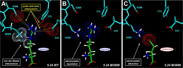
The identification of the chemical structure of an odorant by the vertebrate olfactory system is thought to occur through the combinatorial activity from multiple receptors, each tuned to recognize different chemical features. What are the molecular determinants underlying the selectivity of individual odorant receptors for their cognate ligands? To address this question, we performed molecular modeling and site-directed mutagenesis on the ligand-binding region of two orthologous amino acid odorant receptors belonging to the "C family" of G-protein-coupled receptors in goldfish and zebrafish. We identified the critical ligand-receptor interactions that afford ligand binding as well as selectivity for different amino acids. Moreover, predictions regarding binding pocket structure allowed us to alter, in a predictable manner, the receptor preferences for different ligands. These results reveal how this class of odorant receptor has evolved to accommodate ligands of varying chemical structure and further illuminate the molecular principles underlying ligand recognition and selectivity in this family of chemosensory receptors.


Figures

Figure 1.
Figure 1.
Homology model of receptor 5.24 based on the crystal structure of mGlu1. A, Ribbon diagram of receptor 5.24 extracellular domain from amino acids 28-484. Lobe 1 is depicted in cyan, whereas lobe 2 is shaded magenta. The flexible hinge region is indicated in yellow. The structure is shown in the ligand-bound state in a closed conformation, with an l-arginine molecule present in the binding cleft. The α-carbon of the ligand is toward the left, with the side chain extending toward the right. B, Structural diagram of the proposed binding pocket of receptor 5.24 binding l-arginine. Amino acids proposed to interact with an arginine ligand are indicated by the residue number. Ligand contacts through hydrogen bonds (or ionic interactions in the case of E47 and D388) are depicted by green dashed lines, whereas inter-residue hydrogen bonds are shown as red dashed lines. Side chains of residues S111, S150, E47, and D388 may interact with the ligand via bridging water molecules. Residues D195 (hinge) and K283 have been omitted for clarity. The color scheme and general orientation of the receptor are the same as in A. On the ligand, hydrogens are shown in white, carbons are shown in green, nitrogens are shown in blue, and oxygens are shown in red.
Figure 2.
Figure 2.
Model for arginine binding in receptor 5.24: contribution of residues in the proximal, middle-distal, and far-distal binding pockets. A model of the receptor 5.24 binding pocket bound with l-arginine is shown. This view shows polar interactions with the α-amino and α-carboxylate groups of the ligand in the proximal pocket (S152, T175, Y223, and D309), possible hydrophobic interactions with the backbone of the side chain of the ligand in the middle-distal pocket (S151, A173, K283, and M389), and polar-ionic interactions with the distal guanidinium of the ligand (S111, S150, E47, Y72, D388, S284, S285, and N310). The color scheme is the same as in Figure 1 B, except that residues proposed to form a hydrophobic ring in the middle-distal pocket are highlighted in yellow.
Figure 3.
Figure 3.
Methionine-389 is a critical determinant of ligand selectivity in receptor 5.24. A, Schematic view of selected distal pocket residues (M389, D388, Y72, and E47) involved in contacting the guanidinium moiety of an arginine ligand in the wild-type (WT) receptor. The orientation of this “top-down” view is from above lobe 1 looking down onto the bound ligand, with the proximal pocket toward the bottom and the distal pocket toward the top of the figure. Receptor residues are shaded cyan, with oxygens shown in red. On the ligand, hydrogens are shown in white, carbons are shown in green, nitrogens are shown in blue, and oxygens are shown in red. Proposed intermolecular interactions and distances are indicated with dashed green lines. M389 faces toward the binding pocket, possibly making van der Waals contacts with the n-aliphatic side chain of the docked arginine (Fig. 4 A). B, Predicted interaction of K389 in the receptor 5.24 M389K mutant with bound glutamate. In this model of the mutant binding pocket, the amino group of the side chain of K389 (nitrogen atom shown in blue) can make a direct ionic interaction with the distal carboxylate of the bound glutamate (distances indicated). C, Representative dose-response curves for wild-type and M389K receptor 5.24. HEK293 cells expressing wild-type or mutant receptor were exposed to arginine (blue lines) or glutamate (red lines), and receptor activation was measured by calcium imaging (see Materials and Methods). Note that the wild-type receptor prefers arginine to glutamate, whereas this selectivity is inverted in the M389K mutant. EC50 values for the curves shown in this panel are as follows. Wild type: 0.82 μm arginine, 29.6 μm glutamate; M389K mutant: 24.9 μm arginine, 2.1 μm glutamate.
Figure 4.
Figure 4.
Models of receptor-ligand interactions in wild-type (WT) and M389K mutant receptor 5.24. Proposed hydrophobic and electrostatic interactions between distal binding pocket residues and docked ligands are shown for wild-type and M389K mutant receptor 5.24. The top-down orientation of these views is the same as in Figure 3, A and B. Van der Waals spheres for selected atoms are displayed (white, hydrogen; blue, nitrogen; red, oxygen). A, For arginine docked in the wild-type binding pocket, M389 makes van der Waals contacts with the methylene backbone of the n-aliphatic side chain of the ligand. This may serve to hold the side chain of the ligand in an extended conformation, such that the distal guanidinium group can participate in favorable polar and ionic interactions with D388, Y72, and E47 in the far-distal pocket. B, In the M389K mutant, electrostatic repulsion occurs between the distal amino group of K389 and the guanidinium of the arginine ligand. This unfavorable interaction is shown here for illustrative purposes, although in reality the repulsion probably prevents binding in this conformation. C, The lysine residue introduced into the binding pocket of the M389K mutant is positioned to form a favorable electrostatic interaction (a salt bridge) with the distal carboxylate of the glutamate ligand. Consistent with this model, the M389K mutant shows a marked increase in selectivity for glutamate over arginine.
Figure 5.
Figure 5.
Zebrafish receptor ZO6 is a glutamate receptor and can be rationally retuned by the K386M mutation. A, View of the receptor ZO6 binding pocket looking down onto glutamate docked in the ligand-binding pocket from the perspective of lobe 1. Distal pocket residues K386, D385, Y70, and E45 (corresponding, respectively, to M389, D388, Y72, and E47 in receptor 5.24) are highlighted. The amino group of the K386 side chain can make a direct ionic interaction with the distal carboxylate of the bound glutamate (distances indicated). B, Representative dose-response curves for wild-type (WT) and K386M receptor ZO6. HEK293 cells expressing wild-type or mutant receptor were exposed to arginine (blue lines) or glutamate (red lines), and receptor activation was measured by calcium imaging (see Materials and Methods). Note that the wild-type zebrafish receptor prefers glutamate to arginine, whereas the K386M mutant is rendered nonselective for glutamate and arginine. EC50 values for the curves shown in this panel are as follows. Wild type: 671 μm arginine, 7.1 μm glutamate; K386M mutant: 80.8 μm arginine, 126 μm glutamate.

References

    1. Araneda RC, Kini AD, Firestein S (2000) The molecular receptive range of an odorant receptor. Nat Neurosci 3: 1248-1255. - PubMed
    1. Armstrong N, Sun Y, Chen GQ, Gouaux E (1998) Structure of a glutamate-receptor ligand-binding core in complex with kainate. Nature 395: 913-917. - PubMed
    1. Barth AL, Dugas JC, Ngai J (1997) Noncoordinate expression of odorant receptor genes tightly linked in the zebrafish genome. Neuron 19: 359-369. - PubMed
    1. Bertrand HO, Bessis AS, Pin JP, Acher FC (2002) Common and selective molecular determinants involved in metabotropic glutamate receptor agonist activity. J Med Chem 45: 3171-3183. - PubMed
    1. Bessis AS, Bertrand HO, Galvez T, De Colle C, Pin JP, Acher F (2000) Three-dimensional model of the extracellular domain of the type 4a metabotropic glutamate receptor: new insights into the activation process. Protein Sci 9: 2200-2209. - PMC - PubMed

Publication types

MeSH terms

Associated data