. 2004 Nov 10;24(45):10248-59.
doi: 10.1523/JNEUROSCI.0546-04.2004.

Regulation of the NMDA receptor complex and trafficking by activity-dependent phosphorylation of the NR2B subunit PDZ ligand

Affiliations

Regulation of the NMDA receptor complex and trafficking by activity-dependent phosphorylation of the NR2B subunit PDZ ligand

Hee Jung Chung et al. J Neurosci. .

Abstract

Interactions between NMDA receptors (NMDARs) and the PDZ [postsynaptic density-95 (PSD-95)/Discs large/zona occludens-1] domains of PSD-95/SAP90 (synapse-associated protein with a molecular weight of 90 kDa) family proteins play important roles in the synaptic targeting and signaling of NMDARs. However, little is known about the mechanisms that regulate these PDZ domain-mediated interactions. Here we show that casein kinase II (CK2) phosphorylates the serine residue (Ser1480) within the C-terminal PDZ ligand (IESDV) of the NR2B subunit of NMDAR in vitro and in vivo. Phosphorylation of Ser1480 disrupts the interaction of NR2B with the PDZ domains of PSD-95 and SAP102 and decreases surface NR2B expression in neurons. Interestingly, activity of the NMDAR and Ca2+/calmodulin-dependent protein kinase II regulates CK2 phosphorylation of Ser1480. Furthermore, CK2 colocalizes with NR1 and PSD-95 at synaptic sites. These results indicate that activity-dependent CK2 phosphorylation of the NR2B PDZ ligand regulates the interaction of NMDAR with PSD-95/SAP90 family proteins as well as surface NMDAR expression and may be a critical mechanism for modulating excitatory synaptic function and plasticity.


Figures
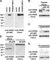
Figure 1.
Figure 1.
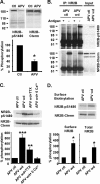
The C-terminal PDZ ligand of NR2B is phosphorylated at Ser1480 in vivo. A-C, Characterization of the phosphorylation site-specific anti-NR2B-pS1480 antibody using immunoblots of HEK293T cell crude membranes expressing NR2B. After immunoblotting with anti-NR2B-pS1480 antibody, the blot was stripped and reblotted with phosphorylation-independent anti-NR2B C-terminal antibody. A, Anti-NR2B-pS1480 antibody recognized NR2B in cells transfected with wild-type NR2B (NR2B) but not empty vector (mock) or the mutant NR2B (NR2BS1480), in which Ser1480 was mutated to alanine. B, Anti-NR2B-pS1480 antibody did not recognize NR2B when anti-NR2B-pS1480 antibody was preincubated with Ser1480-phosphorylated NR2B C-terminal peptide (p2B) but not unphosphorylated peptide (2B). C, Anti-NR2B-pS1480 antibody no longer recognized NR2B when the blot was pretreated with λ-phosphatase before immunoblotting. D, E, Western blot for NR2B-pS1480 in rat brain membrane homogenates. D, Anti-NR2B-pS1480 antibody recognized NR2B in rat brain. Preincubation of anti-NR2B-pS1480 antibody with Ser1480-phosphorylated NR2B C-terminal peptide (p2B) but not unphosphorylated peptide (2B) blocked NR2B recognition by anti-NR2B-pS1480 antibody. E, Anti-NR2B-pS1480 antibody no longer recognized NR2B when the blot was pretreated with λ-phosphatase before immunoblotting.
Figure 2.
Figure 2.
NMDAR activity regulates phosphorylation of the NR2B PDZ ligand. Quantitative immunoblot analysis with anti-NR2B-pS1480 and anti-NR2B C-terminal antibodies was performed on the crude membranes of cultured cortical neurons. A, Chronic APV treatment of cultured cortical neurons decreased phosphorylation of NR2B Ser1480 (APV; n = 4; t test; *p < 0.05) compared with the control neurons maintained in APV-free medium (Ctl), without affecting the level of NR2B expression. B-D, Synaptic NMDARs were activated by removing APV for 15 min from the chronic APV-treated neurons (APV wd). The APV control neurons were maintained in APV (APV ctl). B, IP of NR2B subunits using anti-NR2B C-terminal antibody. Synaptic activation of NMDARs by APV withdrawal increased phosphorylation of NR2B Ser1480. C, APV withdrawal significantly increased phosphorylation of NR2B Ser1480 compared with the APV control (n = 6; t test; ***p < 0.001). This increase was partially blocked by 1 μm TTX (APV wd+TTX; n = 4; t test; **p < 0.01) and was abolished by APV withdrawal without calcium (APV wd+0 mm Ca2+; n = 4; t test; p > 0.05). D, APV withdrawal significantly increased Ser1480 phosphorylation of surface biotinylated NR2B subunits compared with APV control (APV ctl; n = 4; t test; *p < 0.05).
Figure 3.
Figure 3.
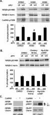
CaMKII, but not PKC or PKA, regulates Ser1480 phosphorylation of NR2B during synaptic NMDAR activation. Quantitative immunoblot analysis with anti-NR2B-pS1480 and anti-NR2B C-terminal antibodies was performed on the crude membranes of cultured cortical neurons. A, The chronic APV-treated cortical neurons were incubated with DMSO (0.1% v/v), KN93 (5-10 μm; CaMKII inhibitor), or KN92 (5-10 μm; inactive analog of KN93) for 1 hr before APV withdrawal. Anti-CaMKII-pT286 antibody (CaMKII-pT286) detected autophosphorylation of CaMKII at Thr286. KN93 completely blocked CaMKII activation and partially blocked phosphorylation of NR2B Ser1480 induced by APV withdrawal (APV wd plus DMSO and APV wd plus KN93; n = 4; t test; *p < 0.05). B, The chronic APV-treated cortical neurons were incubated with DMSO (0.1% v/v), chelerythrine (5 μm; PKC inhibitor), or RP-8-Br-cAMP (10 μm; PKA inhibitor) for 1 hr before APV withdrawal. Chelerythrine and RP-8-Br-cAMP had no effect on APV withdrawal-induced phosphorylation of NR2B Ser1480 (APV wd plus DMSO and APV wd plus chelerythrine or RP-8-Br-cAMP; n = 3; t test; p > 0.05). C, In vitro phosphorylation reactions of GST fusion proteins of the NR2B C-terminal tail and GluR1 C-terminal tail were performed with CaMKII (50 U). Phosphorylation of NR2B Ser1480 and GluR1 Ser831 (a previously characterized CaMKII site) was detected by immunoblotting with anti-NR2B-pS1480 and anti-GluR1-pS831 antibodies, respectively. CaMKII directly phosphorylated GluR1 Ser831 but not NR2B Ser1480. The asterisk indicates the C-terminally truncated GluR1 fusion protein, which is recognized by anti-GluR1-pS831 antibody but not by anti-GluR1 C-terminal antibody.
Figure 4.
Figure 4.
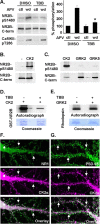
CK2 functions downstream of CaMKII during synaptic NMDAR activation and directly phosphorylates Ser1480. A, The chronic APV-treated cortical neurons were incubated with DMSO (0.1% v/v) or TBB (10-20 μm; CK2 inhibitor) for 1 hr before APV withdrawal. Quantitative immunoblot analysis with anti-NR2B-pS1480 and anti-NR2B C-terminal antibodies revealed that TBB completely blocked APV withdrawal-induced phosphorylation of NR2B Ser1480 (APV wd plus DMSO and APV wd plus TBB; n = 4; t test; *p < 0.05) but not autophosphorylation of CaMKII at Thr286. B, C, Immunoblot analysis of in vitro phosphorylation reactions of GST fusion proteins of the NR2B C-terminal tail by CK2 (20 U; B) and CK2, GRK2, and GRK5 (0.4 μg each; C). B, CK2 directly phosphorylated NR2B Ser1480. C, GRK2 and GRK5 phosphorylated NR2B Ser1480 very weakly compared with the same amount of CK2. D, Autoradiograph and Coomassie blue staining of an SDS-PAGE gel containing in vitro phosphorylation reactions of the GST fusion proteins of the NR2B C-terminal tail by CK2 (0.4 μg) with or without TBB (20 μm). TBB inhibited CK2 activity. E, Autoradiograph and Coomassie blue staining of an SDS-PAGE gel containing in vitro phosphorylation reactions of rhodopsin by GRK2 (0.4 μg) with or without TBB (20 μm). TBB did not inhibit GRK2 activity. F, Immunostaining of 3-week-old low-density cultured hippocampal neurons with anti-CK2α (CK2α) and anti-NR1 C-terminal antibodies (NR1). CK2α subunits colocalized with NR1 in the dendritic shafts and some spines. G, Immunostaining of 3-week-old low-density cultured hippocampal neurons with anti-CK2α (CK2α) and anti-PSD-95 antibody (PSD-95). CK2α subunits also colocalized with PSD-95 in some dendritic spines.
Figure 5.
Figure 5.
Preincubation of CaMKII with CK2 increases phosphorylation of NR2B Ser1480 by CK2 in vitro. A, Autoradiograph and Coomassie blue staining of an SDS-PAGE gel containing in vitro phosphorylation reactions of purified CK2 holoenzyme (2 μg) with TBB (20 μm) by purified rat brain CaMKII (50 U). CaMKII directly phosphorylated CK2 β-subunits. B, Immunoblot analysis of in vitro phosphorylation reactions of the GST fusion proteins of the NR2BC-terminal tail by CK2 (20U), which was first phosphorylated by CaMKII (50U) in vitro. After CaMKII phosphorylation of CK2, KN93 was added to block CaMKII activity. Preincubation of CaMKII with CK2 overall does not significantly increase CK2 phosphorylation of NR2B Ser1480 compared with CK2 alone. However, the increase was statistically significant at the 6 min time point (n = 4; t test; *p < 0.05).
Figure 6.
Figure 6.
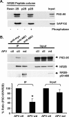
Phosphorylation of the NR2B PDZ ligand disrupts its interaction with SAP102 and PSD-95. A, In vitro binding studies of PSD-95 (top) or SAP102 (bottom) with NR2B peptides immobilized on Affi-Gel resin. Extracts of HEK293T cells expressing PSD-95 or SAP102 were incubated with unphosphorylated (2B), Ser1480-phosphorylated 2B (p2B), or the λ-phosphatase-treated p2B peptides. The bound PSD-95 or SAP102 proteins were detected by immunoblot with anti-PSD-95 antibody or anti-SAP102 antibody, respectively. Both PSD-95 and SAP102 failed to interact with Ser1480-phosphorylated NR2B. B, IP of NMDAR complexes from the chronic APV-treated cortical neurons after APV control (APV ctl) or APV wd using anti-NR1 C-terminal antibodies. Coimmunoprecipitation of NR2B and PSD-95 was analyzed by immunoblot with anti-PSD-95 (PSD-95), NR2B, and anti-NR2B-pS1480 antibodies (NR2B-pS1480). Synaptic activation of NMDAR increased phosphorylation of NR2B Ser1480 and decreased NMDAR interaction with PSD-95 (n = 5; t test; *p < 0.05).
Figure 7.
Figure 7.
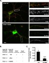
Mutation of Ser1480 to glutamate decreases surface expression of NR2B in neurons. A, Surface immunostaining of GFP-NR2B wild-type and mutant subunit (NR2B S1480E) in which Ser1480 was replaced with glutamate to mimic phosphorylated Ser1480 (left; scale bar, 10 μm). The right panels show higher magnification of the insets in the left panel. B, In the yeast two-hybrid system, PDZ domains of PSD-95 failed to interact with phosphorylation mimic mutant NR2B S1480E and deletion mutant NR2B (NR2B Δ5aa), in which the PDZ ligand was deleted. C, Quantification of surface GFP staining of wild-type NR2B (n = 19) and mutant NR2B S1480E (n = 26) as the ratio of surface GFP intensity over total GFP intensity per unit area (t test; ***p < 0.001).
Figure 8.
Figure 8.
Model for regulation of NMDAR complex and trafficking by phosphorylation of the NR2B PDZ ligand. Synaptic activation of NMDAR stimulates CaMKII and CK2 activity. A, CK2 phosphorylates the PDZ ligand of surface NR2B at Ser1480 and disrupts the interaction between surface NMDAR and the PDZ domains of PSD-95. This leads to destabilization and internalization of surface NMDAR and decoupling of NMDAR from signaling proteins at excitatory synapses. B, CK2 phosphorylates the PDZ ligand of intracellular NR2B at Ser1480 and disrupts the interaction between intracellular NMDAR with the PDZ domains of SAP102. This leads to disruption of SAP102/Sec8-mediated forward trafficking of NMDAR to the cell surface.

Similar articles

Cited by

References

    1. Aoki C, Fujisawa S, Mahadomrongkul V, Shah PJ, Nader K, Erisir A (2003) NMDA receptor blockade in intact adult cortex increases trafficking of NR2A subunits into spines, postsynaptic densities, and axon terminals. Brain Res 963: 139-149. - PubMed
    1. Barria A, Malinow R (2002) Subunit-specific NMDA receptor trafficking to synapses. Neuron 35: 345-353. - PubMed
    1. Bayer KU, De Koninck P, Leonard AS, Hell JW, Schulman H (2001) Interaction with the NMDA receptor locks CaMKII in an active conformation. Nature 411: 801-805. - PubMed
    1. Beattie EC, Carroll RC, Yu X, Morishita W, Yasuda H, von Zastrow M, Malenka RC (2000) Regulation of AMPA receptor endocytosis by a signaling mechanism shared with LTD. Nat Neurosci 3: 1291-1300. - PubMed
    1. Blanquet PR (1998) Neurotrophin-induced activation of casein kinase 2 in rat hippocampal slices. Neuroscience 86: 739-749. - PubMed

Publication types

MeSH terms

Substances