. 2004 Nov 10;24(45):10266-79.
doi: 10.1523/JNEUROSCI.2734-04.2004.

A mutant ataxin-3 putative-cleavage fragment in brains of Machado-Joseph disease patients and transgenic mice is cytotoxic above a critical concentration

Affiliations

A mutant ataxin-3 putative-cleavage fragment in brains of Machado-Joseph disease patients and transgenic mice is cytotoxic above a critical concentration

Daniel Goti et al. J Neurosci. .

Abstract

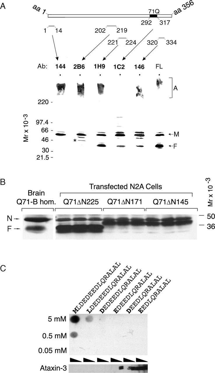
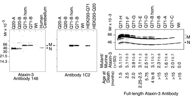
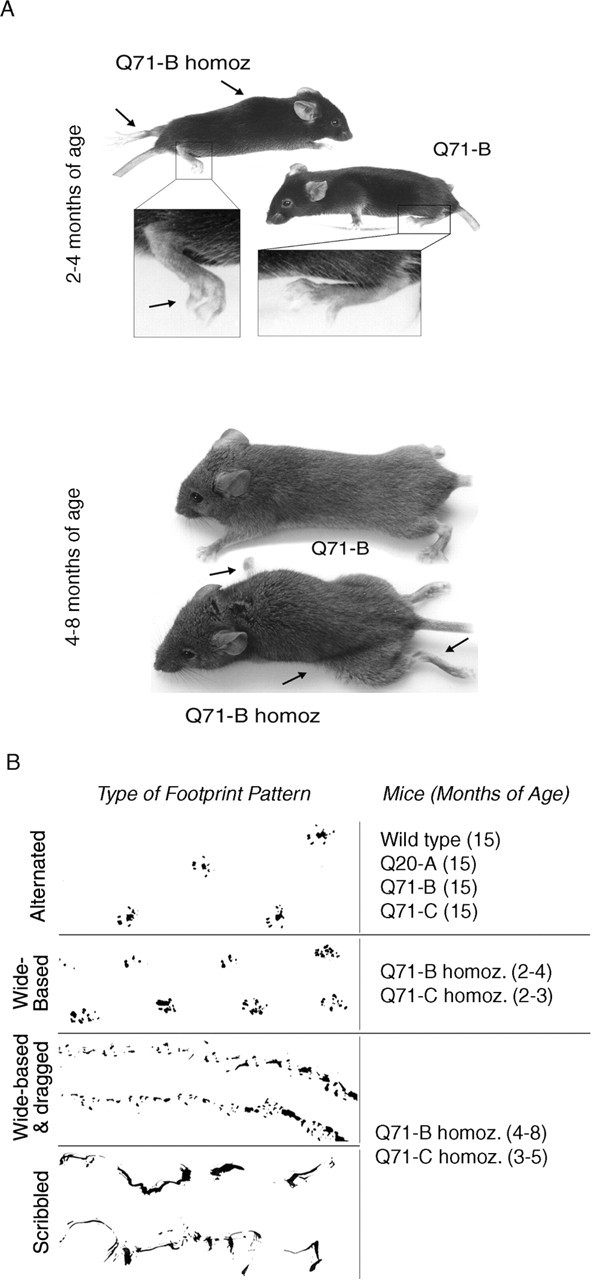
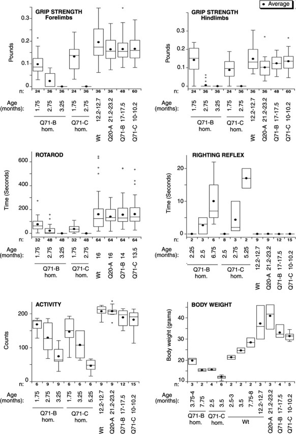
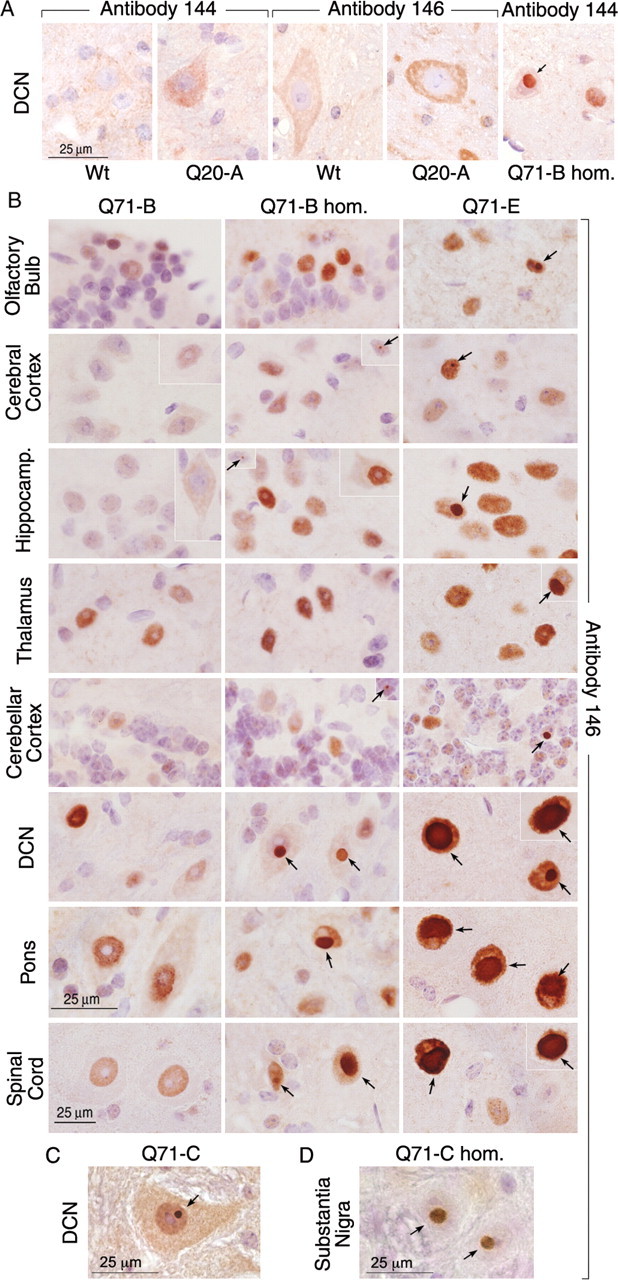
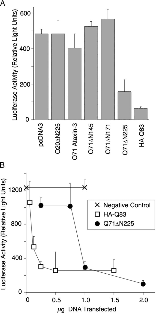
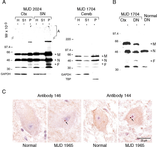
Machado-Joseph disease (MJD) is an inherited neurodegenerative disorder caused by ataxin-3 with a polyglutamine expansion. It is proposed that a toxic cleavage fragment of mutant ataxin-3 alternatively spliced isoform mjd1a triggers neurodegeneration, although this fragment has not yet been detected in the brains of MJD patients or in animal models. We have now generated transgenic mice expressing human mutant (Q71) or normal (Q20) ataxin-3 mjd1a under the control of the mouse prion promoter. Q71 transgenic mice expressing mutant ataxin-3 mjd1a above a critical level developed a phenotype similar to MJD including progressive postural instability, gait and limb ataxia, weight loss, premature death, neuronal intranuclear inclusions, and decreased tyrosine hydroxylase-positive neurons in the substantia nigra (determined by unbiased stereology). Q20 transgenic mice had normal behavior and pathology. Brains from sick Q71 transgenic mice contained an abundant mutant ataxin-3 mjd1a putative-cleavage fragment (Fragment), which was scarce in normal Q71 transgenic mice. Reactivity of the Fragment with a panel of antibodies and comigration with truncations of mutant ataxin-3 revealed that it contained residues C terminal to amino acid 221 to include the polyglutamine expansion. A similar portion of mutant ataxin-3 mjd1a expressed in transfected neuroblastoma cells was toxic above a critical concentration. The Fragment was more abundant in two affected brain regions of MJD patients. Thus, we have developed a murine model for mutant ataxin-3 mjd1a toxicity and identified a putative-cleavage fragment of the disease protein in the brains of these transgenic mice and MJD patients that is cytotoxic above a critical concentration.


Figures

Figure 6.
Figure 6.
Composition of the mutant at axin-3 putative-cleavage fragment. A, Western blot of a brain homogenate of a Q71 transgenic mouse founder (Q71-H) with higher transgene expression levels than Q71-B homozygotes. The blot strips were developed with the ataxin-3 antibodies (Ab) indicated. Antibody 1C2 recognizes preferentially expanded polyglutamines. The antibody labeled here as FL was generated against full-length ataxin-3. The asterisk in the 2B6 blot highlights a nonspecific protein detected with the same intensity in the wild-type mouse brain. Mutant ataxin-3 aggregate (A), full-length form (M), and fragment (F) are highlighted. Normal murine at axin-3 was detected in a longer exposure of the blot (data not shown). B, Western blot of lysates from transiently transfected Neuro-2a cells expressing human mutant at axin-3 mjd1a with a 71 glutamine expansion and missing amino acid residues 1-225 (Q71ΔN225), 1-171 (Q71ΔN171), or 1-145 (Q71ΔN145). Q71-B homozygote brain homogenate (Q71-B hom.) was included as reference to determine which construct was similar in size to the mutant ataxin-3 cleavage fragment. The same amount of protein was analyzed per sample; the lysates were included in triplicate. The blots were developed using an antibody to full-length ataxin-3. Mutant ataxin-3 fragment (F) and normal murine ataxin-3 (N) are highlighted. We note that a higher molecular weight band was detected with all constructs (data not shown), which could be the result of oligomerization resistant to reducing conditions. C, Fine epitope mapping of 1H9 monoclonal antibody was performed using dot blot and competitive assays. Six synthetic polypeptides were designed with the first five amino acids of 221-MLDEDEEDLQRALAL-235 antigenic polypeptide gradually removed. On the dot blot assay (top), increasing amounts of the polypeptides were dotted on nitrocellulose membrane and then immunodetected with 1H9 antibody. For the competitive assay (bottom), 1H9 antibody was preincubated with increasing amounts of the polypeptides (excess of 1, 100, or 1000 molar ratio) before incubation with the Western blot strips containing a protein extract from human lymphoblasts.
Figure 1.
Figure 1.
Transgene expression in the Q71 and Q20 transgenic mouse brain. Western blots of brain homogenates from the indicated transgenic mice [all heterozygous, except for Q71-B homozygotes (hom.)] or wild-type mice (Wt), human cerebellum, or lysates of transfected HEK293 cells expressing the MoPrP constructs (Q71 or Q20) are shown. The same amount of protein was analyzed per sample. The blots were revealed with the indicated ataxin-3 antibodies or antibody 1C2, which preferentially recognizes expanded polyglutamine repeats. The relative migration (Mr) of each molecular weight standard used is indicated. Human mutant ataxin-3 (M) and human or murine normal ataxin-3 (N) are highlighted. The ratio of human mutant over murine ataxin-3 PhosphorImager units is shown under one blot. The results are the average of the values obtained in three similar blots and the corresponding SD (±SD). The ratio for Q71-C homozygotes was not calculated but is expected to be a maximum of double the value shown for their heterozygous parents (Q71-C). Q71 transgenic mice developed the abnormal behavior described in the following figures and had premature death (age at death) or had a normal behavior and were killed at 15 months of age (>15).
Figure 2.
Figure 2.
Appearance and gait of Q71 and Q20 transgenic mice. A, Q71-B homozygotes (Q71-B homoz) 2-4 months of age had a small hunchback, transiently clutched paws, and uncoordinated extension of hindlimbs (arrows). At 4-8 months of age, they had pronounced hunchback with low pelvic elevation and muscle wasting, permanently clutched paws, and uncoordinated movement of extended limbs (arrows). Q71-B heterozygotes (Q71-B) of the same age and sex had the appearance and behavior of wild-type mice. B, The indicated Q71 transgenic mice at the specified ages developed a progressive deteriorating footprint pattern (wide-based at an early stage, wide-based and dragged at a late stage, and scribbled at a later to moribund stage). The remainder of the transgenic mice listed, like wild-type mice, had a normal alternative footprint pattern.
Figure 3.
Figure 3.
Behavioral test and body weight of Q71 and Q20 transgenic mice. For each animal indicated, we determined the following: grip strength, the force (in pounds) needed to release the grip of a rod; Rotarod, the time (in seconds) taken to fall off an accelerating rod; righting reflex, the time (in seconds) required for the mouse to turn its body to a normal position after being placed on its back; activity, the counts detected by a passive infrared activity monitor; and body weight. The values obtained for a given group of animals on different trials on consecutive days were pooled (n = total number of values) and are represented in a box plot. The black circle represents the average, and the line dividing the box represents the median; one-fourth of the data fall between the bottom of the box and the median, and another one-fourth fall between the median and the top of the box. The lines attached to the box extend to thesmallest and the largest data values. Outliers are indicated as small circles and defined as values smaller than the lower quartile minus 1.5 times the interquartile range or larger than the upper quartile plus 1.5 times the interquartile range. hom., Homozygotes; Wt, wild type.
Figure 4.
Figure 4.
Ataxin-3-immunostained neurons from Q71 and Q20 transgenic mice. Paraffin-embedded midsagittal brain and spinal cord sections of the indicated animals were stained with ataxin-3 antibody 146 or 144, as indicated, counterstained with hematoxylin, and analyzed by light microscopy. An inset of an additional image of a neuron is included where necessary, to provide a better representation of the data. The magnification of brain images is the same for all samples and is represented with a bar. The magnification in all spinal cord images is slightly higher and represented by a different length bar. The intranuclear inclusions are highlighted (arrows). A, Images of deep cerebellar nuclei (DCN) neurons of wild-type mice (Wt; 12 months of age), Q20-A transgenic mice (6 months of age), and Q71-Bhomozygotes (Q71-Bhom.; 4 months of age). B, Images of neurons from the indicated brain regions of a 4-month-old Q71-B heterozygote (Q71-B) and Q71-B homozygote (Q71-B hom.) and a 2.2-month-old Q71-E transgenic mouse. C, Image of a deep cerebellar nuclei (DCN) neuron from a Q71-Cheterozygote at 13 months of age. D, Image of substantia nigra neurons of a 2.2-month-old Q71-C homozygote (Q71-C hom.) at 2.75 months of age.
Figure 5.
Figure 5.
Mutant ataxin-3 putative-cleavage fragment in brains of transgenic mice. A, Top and bottom, Western blot of brain homogenates (H) and cytoplasmic (S1) and purified nuclear (P) fractions from wild-type mice (Wt) and the indicated transgenic mice [all heterozygous, except for Q71-B homozygotes (Q71-B hom.)]. The blot was developed with antibody to full-length ataxin-3 (top blot), or as indicated to GAPDH (a cytoplasmic fraction marker) or TBP (a nuclear fraction marker). Bottom, Western blot of brain homogenates from the indicated Q71 transgenic mice and wild-type mice. The right portion of the blot is also shown as an enhanced image (Enhanced). The age at death of the “sick” Q71 transgenic mice is indicated. The “healthy” Q71 transgenic mice had no premature death (>15). Mutant ataxin-3 aggregate (A), full-length form (M), fragment (F), and normal human or murine ataxin-3 (N) are highlighted. B, Western blot of cerebral cortex (Ctx) or cerebellum (Cereb.) homogenates from 4-month-old Q71-B homozygotes (Q71-B hom.). The same amount of protein was analyzed per sample. The blot was developed using the antibody to full-length ataxin-3. Mutant ataxin-3 aggregate (A), full-length form (M), fragment (F), and murine ataxin-3 (N) are highlighted. An enhanced image of the portion of the blot that included the mutant at axin-3 fragment is shown (Fe). C, RT-PCR of brain mRNA from the indicated transgenic and wild-type mice using the indicated pairs of primers. The pairs of primers #1 and #2 are represented under the diagram of the primary structure of human ataxin-3 mjd1acDNA. Thenucleotide number at the 5′ end of each primer is indicated on an arrow. As a control (C), RT-PCR of the same mRNA brain samples was done using primers specific for 18 SrRNA. We did the reaction with or without RT, as indicated. The size markers were Lamda DNA HindIII (first lane) and 1 kb ladder (second lane).
Figure 7.
Figure 7.
Cytotoxicity of truncated mutant ataxin-3. A, Luciferase activity in lysates of Neuro-2a cells transiently cotransfected with the pcDNA3 vector (pcDNA3) or the indicated pcDNA3 constructs and the pRL-SV40-Renilla luciferase plasmid. The constructs used were as follows: (1) human normal ataxin-3 mjd1a with a stretch of 20 glutamines and missing amino acid residues 1-225 (Q20ΔN225); (2) human mutant ataxin-3 mjd1a with a stretch of 71 glutamines (Q71-ataxin-3) or missing amino acid residues 1-145 (Q71ΔN145), 1-171 (Q71ΔN171), or 1-225 (Q71ΔN225); and (3) a stretch of 83 glutamines tagged at the N terminus with an HA epitope and previously generated (HA-Q83). The results are the mean of the values obtained in three experiments with the corresponding SD. B, Luciferase activity in lysates of Neuro-2a cells transiently transfected with 0.2 μg of SV40-Renilla luciferase plasmid by itself (0 μg of DNA transfected, negative control) or together with the indicated quantity of pcDNA3 vector (negative control), HA-Q83, or Q71ΔN225. The results are the mean of the values obtained in three experiments with the corresponding SD. For the 0.25 μg of Q71ΔN225 point, the best two of three values were selected.
Figure 8.
Figure 8.
Mutant ataxin-3 cleavage fragment in the brain of an MJD patient. A, Western blots of homogenates (H) and cytoplasmic (S1) and purified nuclear (P) fractions from frontal cerebral cortex (Ctx), substantia nigra (SN), or cerebellar cortex/dentate nuclei (Cereb) from the indicated MJD heterozygous patients. The same amount of total protein was analyzed per sample, except for SN (1.5 times as much protein was used). The blots were developed using an antibody to full-length ataxin-3 (top blot) or, as indicated, GAPDH (a cytoplasmic fraction marker) or TBP (a nuclear fraction marker). The relative migration (Mr) of each molecular weight standard used is indicated. Mutant ataxin-3 aggregate (A), full-length form (M), fragment (F), and normal ataxin-3 (N) are highlighted. B, Western blot of homogenates from frontal cerebral cortex (Ctx) or dentate nuclei (DN) of MJD heterozygous patient 1704 or normal DN from individual 48,108. The same amount of protein was analyzed per sample. The blot was developed using the full-length ataxin-3 antibody. C, Images of dentate nuclei neurons of the cerebellum from a normal individual or MJD heterozygous patient 1965. Paraffin-embedded sections were stained with ataxin-3 antibody 146 or 144, as indicated, counterstained with hematoxylin, and analyzed by light microscopy. The same magnification was used for all images and is represented with a bar.

References

    1. Berke SJ, Schmied FA, Brunt ER, Ellerby LM, Paulson HL (2004) Caspase-mediated proteolysis of the polyglutamine disease protein ataxin-3. J Neurochem 89: 908-918. - PubMed
    1. Blobel G, Potter VR (1966) Nuclei from rat liver: isolation method that combines purity with high yield. Science 154: 1662-1665. - PubMed
    1. Borchelt DR, Davis J, Fischer M, Lee MK, Slunt HH, Ratovitsky T, Regard J, Copeland NG, Jenkins NA, Sisodia SS, Price DL (1996) A vector for expressing foreign genes in the brains and hearts of transgenic mice. Genet Anal 13: 159-163. - PubMed
    1. Burright EN, Clark HB, Servadio A, Matilla T, Feddersen RM, Yunis WS, Duvick LA, Zoghbi HY, Orr HT (1995) SCA1 transgenic mice: a model for neurodegeneration caused by an expanded CAG trinucleotide repeat. Cell 82: 937-948. - PubMed
    1. Butler R, Leigh PN, McPhaul MJ, Gallo JM (1998) Truncated forms of the androgen receptor are associated with polyglutamine expansion in X-linked spinal and bulbar muscular atrophy. Hum Mol Genet 7: 121-127. - PubMed

Publication types

MeSH terms

LinkOut - more resources